Human corneal endothelial cell sheet

a technology of endothelial cells and corneas, which is applied in the field of human corneal endothelial cells, can solve the problems of reduced cell number, drastic decrease in eyesight, and endothelial cells that cannot grow

Inactive Publication Date: 2015-12-31
NITTA GELATIN INC +1
View PDF2 Cites 9 Cited by
  • Summary
  • Abstract
  • Description
  • Claims
  • Application Information

AI Technical Summary

Benefits of technology

[0036]According to the present invention, a material for corneal endothelial tissue regenerative medicine, which is superior to conventional materials, can be provided.

Problems solved by technology

Corneal endothelial cells do not grow and are not repaired in vivo, and the cell number decreases when they are damaged by trauma in surgery and the like or due to aging.
Bullous keratopathy is a disease wherein corneal stroma develops edema and opacity due to a decrease in the corneal endothelial cell density, thus resulting in a drastic decrease in the eyesight.
On the contrary, corneal transplantation is actually performed in 2500 cases annually and overwhelming shortage of cornea is widespread.
Consequently, patients experiencing obstruction in daily living are forced to wait several years for the transplantation.
However, the patients feel less satisfactory because strong irregular astigmatism, myopia and hyperopia occur after transplantation.
Furthermore, the hospitalization period is long, actual stabilization of the eyesight often takes more than one year, and the eyesight 1.0 is restored less often.
However, the thickness of graft which reaches 150 μm causes optical loss in the interface, and corrected eyesight cannot be sufficiently achieved (corrected eyesight about 0.5).
In view of the importance of uniform lamination of cells on a support, moreover, conception of seeding of cells on a sheet with a curved surface is far from achievable from the common knowledge.

Method used


Image

  • Human corneal endothelial cell sheet
  • Human corneal endothelial cell sheet
  • Human corneal endothelial cell sheet

Examples


example 1

Preparation of 2% Gelatin Solution

[0072]The operation is desirably performed in a clean bench (safety cabinet). All containers and tools used had been sterilized or were of sterilized disposable type. BeMatrix gelatin LS-H powders (Nitta Gelatin Inc., 2.0 g) were weighted and placed in a bottle. Water (100 mL) for injection was added, and the mixture was shaken at room temperature for 1 hr, and dissolved by shaking in a 50° C. thermostatic tank for 30 min. After cooling at room temperature, the mixture was preserved at 4° C.

example 2

Preparation of Curved Gelatin Sheet

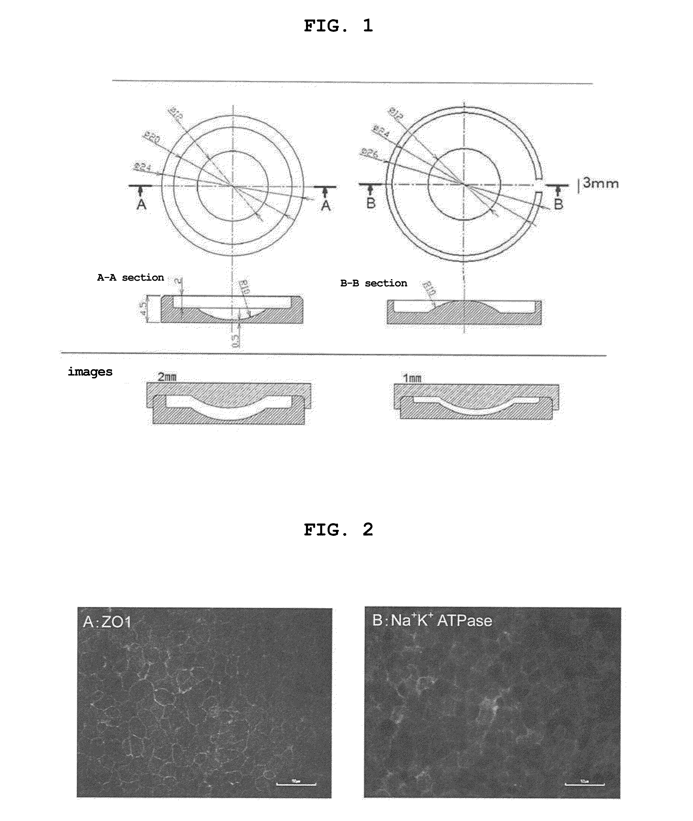
[0073]2% gelatin preserved at 4° C. was dissolved in a thermostatic tank at 37° C. A lid of the Teflon (registered trade mark) template having a curved surface, which was prepared by designing as shown in FIG. 1, was removed, the 2% gelatin solution (600 μL) was poured into the concave surface and spread all over the surface. The lid was placed back thereon, and the gelatin solution was cooled at 4° C. for 1 day to allow for gelation. The lid of the template was slowly opened with tweezers, the gelled gelatin was dried in a clean bench (safety cabinet) for 2 days. After drying, the gelatin in a sheet shape was slowly detached from the template, and crosslinked in a vacuum drying apparatus at 140° C. for 72 hr. As a comparison object, a flat gelatin sheet was prepared in the same manner using a flat template. The gelatin sheet after the crosslinking treatment was preserved at −30° C.

example 3

Adhesion of Gelatin Sheet

[0074]Water (5 mL) for injection was poured into a 35 mm dish, and a curved gelatin sheet was allowed to swell in advance for one day. An insert with a membrane was taken out from Corning Inc. transwell (#3801), and the membrane was removed from the insert with tweezers. Water (5 mL) for injection was poured into a 35 mm dish, and the curved gelatin sheet swollen with water was floated thereon with the convex surface facing upward. A ring after removal of the membrane was placed under the curved gelatin sheet, the circumference of the ring and that of the sheet were matched and the water for injection was extracted. Wrinkles on the sheet were removed with tweezers, and the sheet was air-dried in a clean bench (safety cabinet). The ring and the curved sheet were immobilized with an O-ring, set in the transwell, and swollen with a growth medium.


PUM

PropertyMeasurementUnit
thicknessaaaaaaaaaa
thicknessaaaaaaaaaa
thicknessaaaaaaaaaa

Abstract

The present invention provides a human corneal endothelial cell sheet having a curvature fitting a human corneal endothelial surface, particularly, the human corneal endothelial cell sheet supported by a cell support having a curvature fitting a human corneal endothelial surface, and a production method of a human corneal endothelial cell sheet having a curvature fitting a human corneal endothelial surface, which includes the following steps:(1) a step of gelling a gelatin aqueous solution poured into a template having a curvature fitting a human corneal endothelial surface;(2) a step of forming a sheet-like gelatin by drying the gelled gelatin obtained in (1);(3) a step of obtaining a gelatin sheet by thermally crosslinking the sheet-like gelatin obtained in (2);(4) a step of seeding human corneal endothelial cells on the gelatin sheet obtained in (3) to form a human corneal endothelial cell layer.

Description

TECHNICAL FIELD [0001]The present invention relates to the development of a human corneal endothelial cell sheet and an effective corneal endothelial cell transplantation therapy using same.BACKGROUND ART [0002]Corneal endothelium consists of a single layer of cells constituting the innermost layer of cornea, and plays the most important role in the maintenance of the transparency of cornea, namely, eyesight by its barrier function and pumping function. Corneal endothelial cells do not grow and are not repaired in vivo, and the cell number decreases when they are damaged by trauma in surgery and the like or due to aging. Bullous keratopathy is a disease wherein corneal stroma develops edema and opacity due to a decrease in the corneal endothelial cell density, thus resulting in a drastic decrease in the eyesight. An effective treatment method is corneal transplantation alone. The greatest number of patients that undergo corneal transplantation suffer from bullous keratopathy (about...

Claims


Application Information

Patent Timeline
Patent Type & Authority Applications(United States)
IPC IPC(8): A61L27/38A61L27/34A61L27/54A61L27/36A61L27/22
CPCA61L27/3641C12N2535/00A61L27/222A61L27/24A61L27/34A61L27/3808A61L2430/16C12N5/0621C12N2537/10A61L27/54A61L27/3691A61L2300/64A61L27/22C12N2533/52C12N2533/54C08L89/06A61P27/02A61P43/00
Inventor YAMAGAMI, SATORUSHIMA, NOBUYUKIYAMAUCHI, MIWAYAMAGUCHI, MASAHIROHIRAOKA, YOSUKE
Owner NITTA GELATIN INC
Why Eureka
Try Eureka