

Therapeutic drug for diseases related to endoplasmic reticulum cell death in corneal endothelium
a technology of endoplasmic reticulum cells and therapeutic drugs, which is applied in the direction of antibody medical ingredients, peptide/protein ingredients, and metabolic disorders, etc., can solve the problem that human corneal endothelial cells have very limited regeneration ability
- Summary
- Abstract
- Description
- Claims
- Application Information
AI Technical Summary
Benefits of technology
Problems solved by technology
Method used
Image

Examples
examples
[0105]Hereinafter, examples of the present invention are disclosed. Biological samples or the like, when applicable, were handled in compliance with the standards enacted by the Ministry of Health, Labour and Welfare, Ministry of Education, Culture, Sports, Science and Technology, or the like and, if applicable, based on the Helsinki Declaration or ethical codes prepared based thereon. For the donation of eyes used for the research, consent was obtained from close relatives of all the deceased donors. The present research was approved by the ethics committee or a corresponding body of the University of Erlangen and SightLife™ (Seattle, Wash.) eye bank.
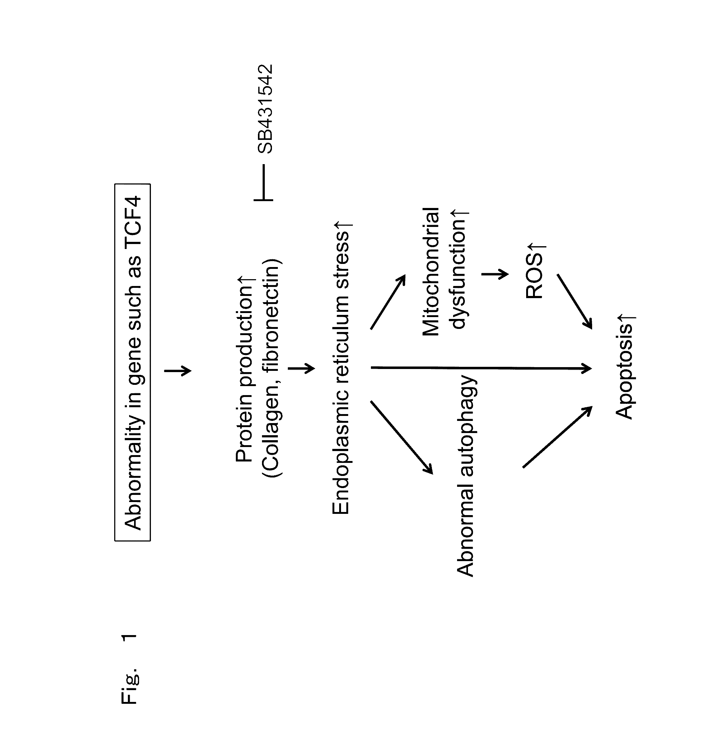
[0106]Fuchs' endothelial corneal dystrophy leads to the death of corneal endothelial cells. When the remaining corneal endothelial cells cannot compensate for the lost pumping function or barrier function, transparency of a cornea cannot be maintained, resulting blindness due to corneal opacity. Further, corneal endothelial cells of a ...
preparation example 2
( Confirmation of Normal Functioning of Immobilized Corneal Endothelial Cell Line (iFECD))
[0112]Normal functioning of an immobilized corneal endothelial cell line (iFECD) was confirmed in the present Example.
(Immunostaining by Na+ / K+-ATPase and ZO-1)
[0113]First, immunostaining was performed with Na+ / K+-ATPase and ZO-1 to confirm normal functioning of an immobilized corneal endothelial cell line (iFECD). This is for examining the functions of corneal endothelial cells, i.e., pumping and barrier functions. Na+ / K+-ATPase and ZO-1 indicates normal pumping and barrier functions of corneal endothelial cells, respectively. The technology is as follows.
[0114](Method of Observing Cells with Staining or the Like (Histological Test))
[0115]Cells were observed with a phase difference microscope. After cells were immobilized, immunostaining was applied with ZO-1 and Na+ / K+-ATPase as function associated markers for observation with a fluorescent microscope. For a tissue staining test, cultured cel...
example 1
Morphological Observation of Endoplasmic Reticulum and Mitochondria in Fuchs' Endothelial Corneal Dystrophy
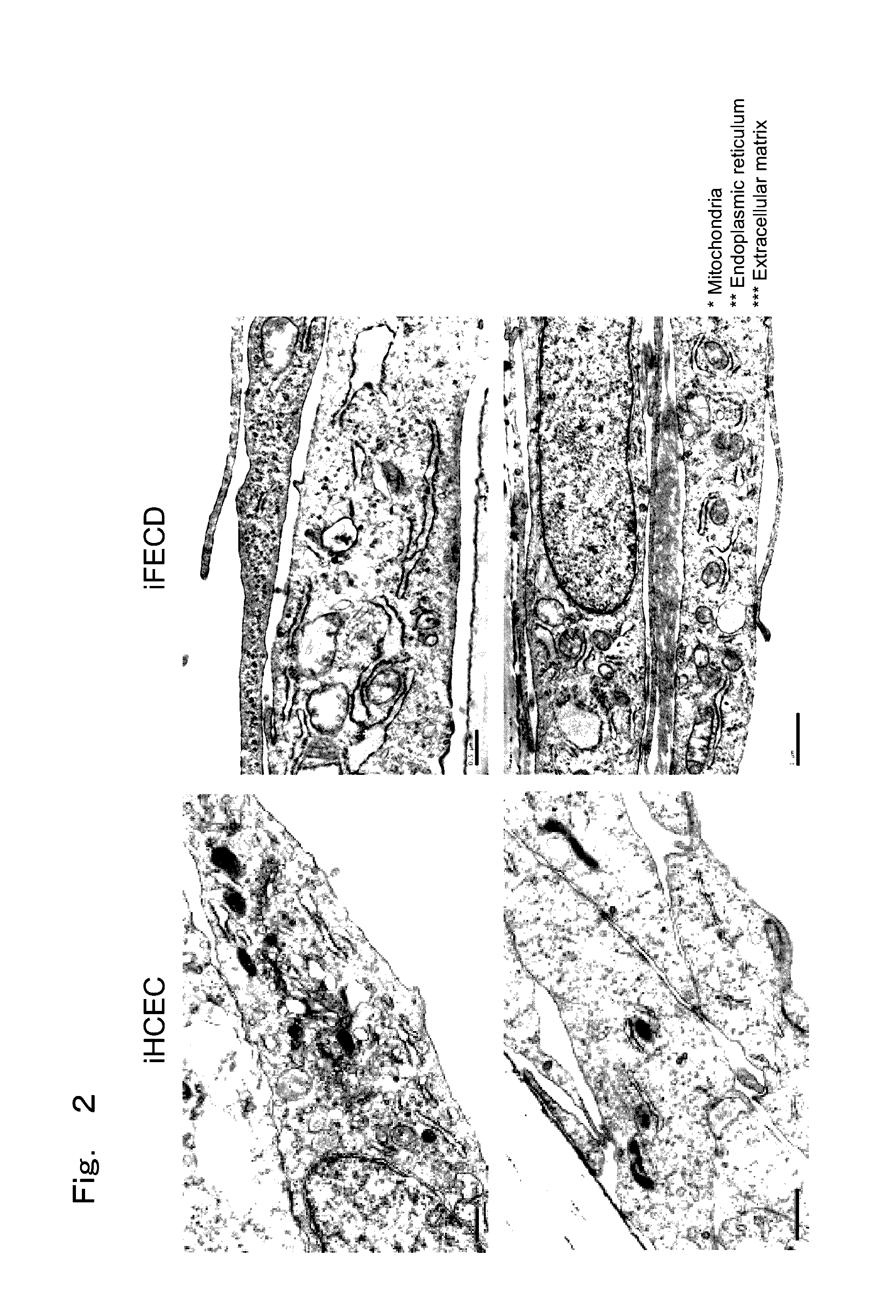
[0119]The present Example observed whether morphological abnormality is observed in endoplasmic reticulum and mitochondria in cells with Fuchs' endothelial corneal dystrophy model prepared in the above-described Preparation Example as a representative example in order to study endoplasmic reticulum stress.
(Observation of Organelles by Electron Microscope)
[0120]Cells were then observed with an electron microscope to confirm elevation in ECM production and ER stress. As a pre-immobilization, a transwell seeded with cells was immobilized for three hours at room temperature with 2.5% glutaraldehyde with a pH 7.2 diluted with 0.1 M sodium cacodylate and washed three times with 0.1 M sodium cacodylate. Then, as post immobilization, it was immobilized for 1 hour at room temperature with 1% osmic acid diluted with 0.1 M sodium cacodylate and washed three times with distilled water. In ...
PUM
| Property | Measurement | Unit |
|---|---|---|
| Stress optical coefficient | aaaaa | aaaaa |
Abstract
Description
Claims
Application Information