Corneal posterior lamella and use thereof

A cornea and posterior plate technology, applied in the field of medical materials, can solve the problems of lack of observation results, shedding of offspring cells, tumorigenesis, etc., and achieve the effect of overcoming low proliferation ability and strong proliferation ability

Inactive Publication Date: 2010-06-23
SHANGHAI NINTH PEOPLES HOSPITAL AFFILIATED TO SHANGHAI JIAO TONG UNIV SCHOOL OF MEDICINE
View PDF0 Cites 9 Cited by
  • Summary
  • Abstract
  • Description
  • Claims
  • Application Information

AI Technical Summary

Benefits of technology

This patented technology allows scientists studying how human eye's inner layers work better by isolating specific types or groups of cells called anterior corneallary epitheliocytes (ACE) before they start growing into connective tissues like collagen fibers inside the stroma layer on the back surface of an eyeball. These ACEs were isolated during embryonic development but could still grow even after birth due to their own growth potential and autonomy. They had many important applications such as creating artificial eyes without relying solely upon animal models, developing new treatments for diseases caused by gene defects affecting vision, testing drug effectiveness through monitoring changes overtime, simulating physical properties of natural ocular media, making prosthetic implants more durable, etc., while avoiding problems associated with current methods involving living subjects.

Problems solved by technology

This patents discusses different types of eye implants called ocular prosthesive graft (OPG) tubule plugging method developed during this patented process. These methods involve transferring autologous crystalline cells grown inside the recipient' s own eyes without causing any complications like blood clots. Additionally, other techniques involving inducement of specific type of organs within the host may improve their functionality compared to existing procedures.

Method used


Image

  • Corneal posterior lamella and use thereof
  • Corneal posterior lamella and use thereof
  • Corneal posterior lamella and use thereof

Examples


Embodiment

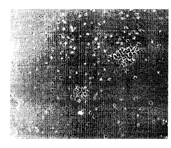
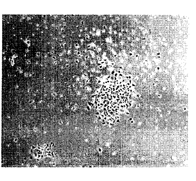
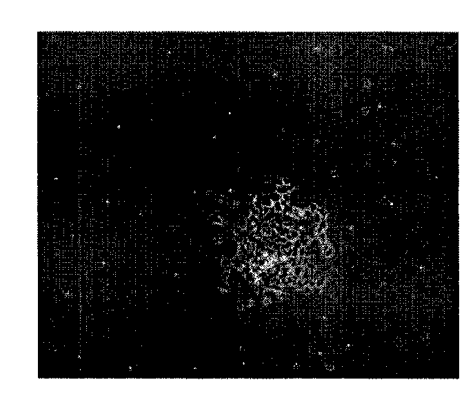
[0030] 1. Isolation and culture of cat corneal endothelial precursor cells

[0031] Put the cat's eyeballs into a sterilized dish after being washed with PBS. Carefully separate the conjunctival tissue, carefully scrape off the epithelium, cut the corneal tissue 1 mm outside the limbus, and place it in DMEM / F12 (1:1) culture solution. Gently scrape off the corneal epithelial cells with a razor blade, and carefully peel off the Descemet's membrane containing endothelial cells with ophthalmic forceps. Cut the Descemet membrane repeatedly, add 0.2% composite collagenase (the volume ratio of Descemet membrane tissue block to collagenase is about 1:5, and the volume ratio of tissue block to collagenase is 1:5, which can achieve the goal of not wasting collagenase , less damage to cells, and can effectively digest the Descemet membrane, shortening the operating time. The volume ratio of tissue block to collagenase is lower than 1:5, and the amount of collagenase is relatively large...


PUM


Abstract

The invention relates to a corneal posterior lamella. The corneal posterior lamella is prepared by the following method: (a) separating and culturing cat corneal endothelial precursor cells for obtaining corneal endothelial precursor cells; (b) preparing a porcine corneal acellular matrix and obtaining the acellular matrix; and (c) constructing the corneal posterior lamella. The invention further provides a use of using the corneal posterior lamella as a donor for corneal transplantation. The structure of the tissue engineering corneal posterior lamella constructed by the corneal endothelial precursor cells is similar to the structure of the normal corneal posterior lamella. The construction of the corneal posterior lamella by taking the corneal endothelial precursor cells as seeds can provide sufficient cell sources, overcome the difficult problem of low proliferation capacity of the corneal endothelial cells and simultaneously solve the problems of cell transformation and safety.

Description


Claims


Application Information

Patent Timeline
Owner SHANGHAI NINTH PEOPLES HOSPITAL AFFILIATED TO SHANGHAI JIAO TONG UNIV SCHOOL OF MEDICINE
Why Eureka
Try Eureka