
Sheet for corneal transplants

a sheet and corneal technology, applied in the field of sheet for corneal transplants, can solve the problems of corneal endothelial disease such as corneal opacity and bullous keratopathy, reduce the number of transplanted cells, and insufficient water discharge, etc., to achieve stable transplantation results, appropriate strength, and easy handling of cells during transplantation

Inactive Publication Date: 2012-11-08
OSAKA UNIV
View PDF7 Cites 9 Cited by
  • Summary
  • Abstract
  • Description
  • Claims
  • Application Information

AI Technical Summary

Benefits of technology

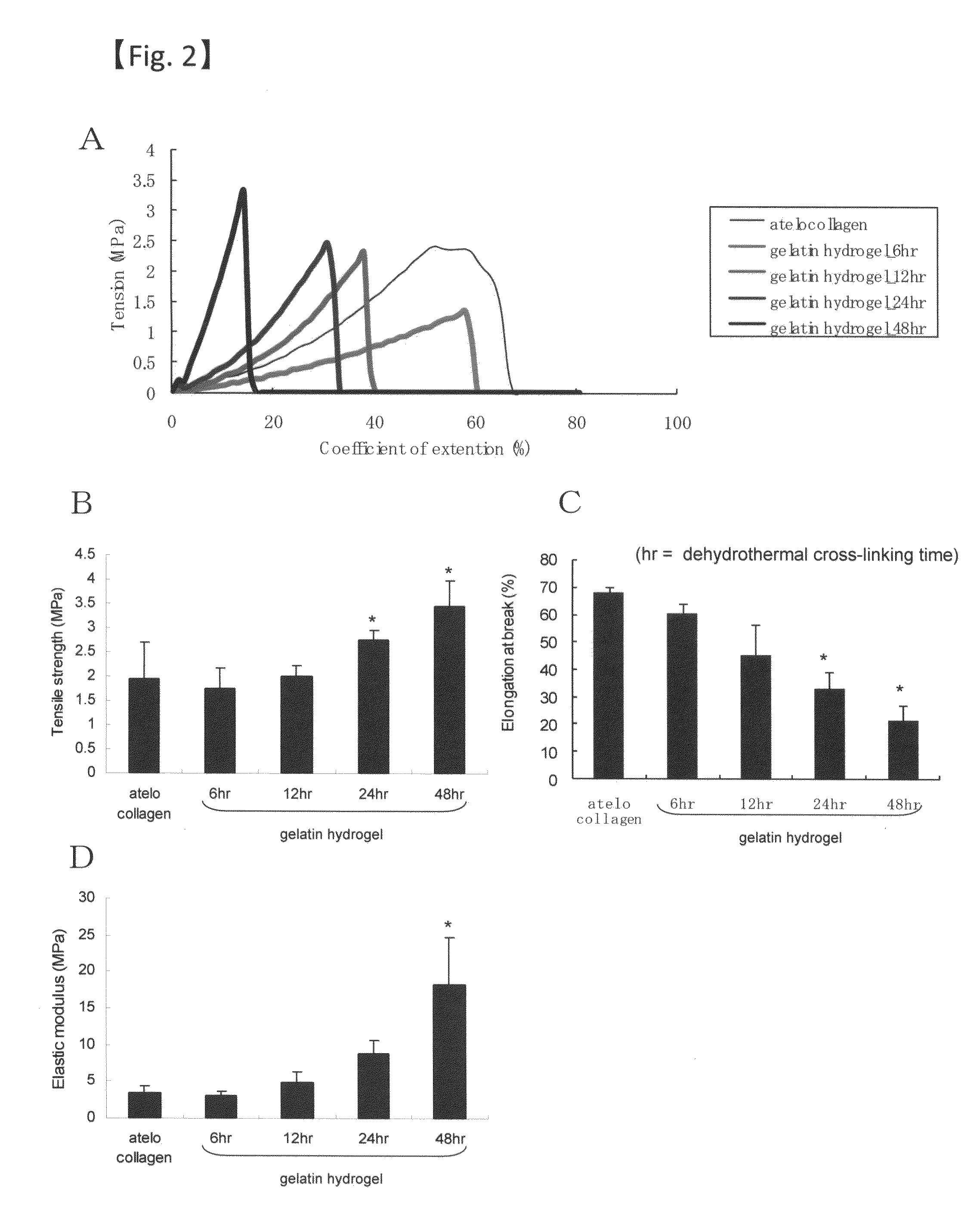
[0027]Because the cultured corneal endothelial cells form a monolayer, handling of the cells during transplantation is difficult. Thus, a strong, transparent carrier is needed to achieve stable transplantation results. The gelatin hydrogel used in the present invention has appropriate strength and excellent transparency, and when used as a carrier for transplantation, it will remarkably improve the strength and operability of the sheet for corneal transplants. Also, because the gelatin hydrogel is degraded in vivo and then disappears, there is no concern that this gelatin hydrogel may cause damage to the corneal endothelium, and further, the corneal transparency is recovered in the early post-transplantation period. Further, because gelatin hydrogels have been already used as a clinical material, the sheet for corneal transplants of the present invention using a gelatin hydrogel can be safely used for clinical application.

Problems solved by technology

Reduction in the number of corneal endothelial cells leads to insufficient water discharge, causing corneal endothelial disease such as corneal opacity and bullous keratopathy.
Conventionally, penetrating keratoplasty has been performed for corneal endothelial disease; however, there are problems of absolute shortage of donors and post-transplant rejection reaction.
However, this method cannot be a radical solution to the problems of rejection reaction and donor shortage.
However, normally, cultured corneal endothelial cells form a monolayer and the intercellular connection is not very strong; therefore, direct use of the cultured corneal endothelial cells for transplantation is difficult in terms of operability and strength.
Also, although cultured cells are normally detached and collected from culture containers by treatment with an enzyme such as trypsin or with chemicals, there are problems that such treatment would disrupt the desmosome structure, causing drastic reduction in the engraftment, strength, and function of the transplanted cell sheet.
However, because no carrier is used in this method, handling of the harvested cultured corneal endothelial cells is difficult, posing a problem for practical clinical application.
However, atelocollagen has poor bioadhesive and biodegradable properties, and there is a report that it exfoliates from the endothelial surface after transplantation and remains in the anterior chamber.
Thus, there is a concern that this might stimulate endothelial cells, consequently causing a reduction in the cell density.
However, the gelatin disk is as thick as about 800 μm and requires a long time for biodegradation, and also, there is a concern that degradation might induce inflammation.
However, the use of a gelatin hydrogel in corneal cell culture as a carrier for corneal endothelial transplantation has been unknown to date.

Method used


Image

  • Sheet for corneal transplants
  • Sheet for corneal transplants
  • Sheet for corneal transplants

Examples


example 1

Preparation of the Gelatin Hydrogel Sheet

[0080]Collagen was extracted from pig skin or cattle bone by alkali treatment (molecular weight: 98,000, isoelectric point: 5.0). Then, a 10 WT % aqueous solution of gelatin was adjusted. This solution was poured into a plastic petri dish and left for several days at room temperature to evaporate water, whereby a gelatin sheet was obtained. The gelatin sheet was then subjected to thermal dehydration treatment at 160° C. and 0.01 Torr for 72 hours to chemically cross-link the gelatin molecules. The gelatin sheet thus obtained had a percentage water content of 97% and a thickness of 100 μm. It should be noted that thickness can be adjusted to 30 μm or more depending on the purpose.

(1) Transparency

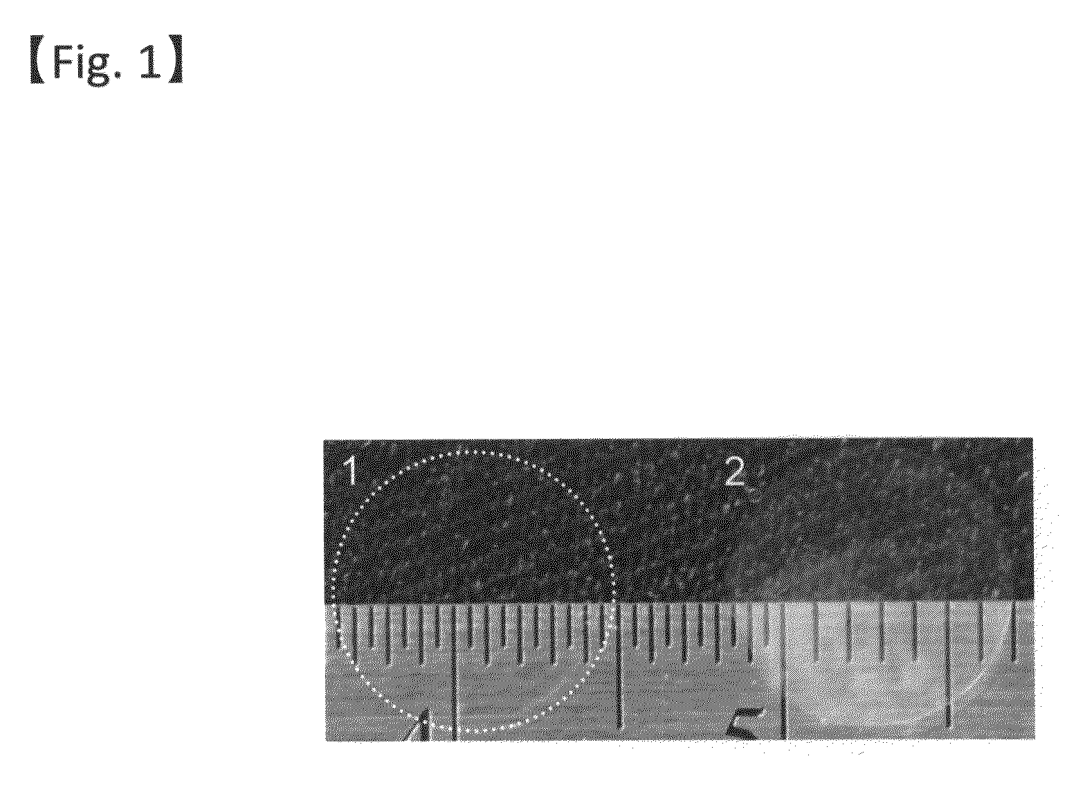
[0081]The gelatin hydrogel sheet prepared as above (48 hours of cross-linking) and an atelocollagen sheet (KOKEN CO., LTD.) were visually compared for transparency (FIG. 1). Further, the light transmittance was compared among the gelatin hydrogel sheet...

example 2

Cell Culture on the Gelatin Hydrogel Sheet

1. Preparation of Corneal Endothelial Cells

[0087]Corneoscleral buttons were prepared from the rabbit eye and the Descemet's membrane was detached. The Descemet's membrane was incubated with 0.25% Trypsin-EDTA at 37° C. for 10 minutes to isolate cells. To the cells, a DMEM medium (low glucose, Nikken Bio Medical Laboratory) containing 2 ng / ml bFGF (R&D systems, Inc.) and 10% serum (Japan bio serum) was added, followed by centrifugation at 300 g for five minutes. The supernatant was aspirated and the remaining cell aggregate was suspended in the same medium.

2. Study on the Corneal Endothelial Cell Culture and the Coating Conditions

[0088]The gelatin hydrogel sheet prepared in Example 1 was coated with type I or type IV collagen (Nitta Gelatin, Inc.). As the coating method, a method of directly applying a 3.0 mg / ml collagen stock solution (pH 3.0) on the gelatin hydrogel sheet prepared in Example 1 with a cell scraper and the like and a method o...

example 3

Transplantation of the Gelatin Hydrogel Sheet into the Rabbit Anterior Chamber (Study on Biodegradability)

[0093]Following cataract extraction from the rabbit eye (phacoemulsification), the Descemet's membrane was detached. Then, a gelatin hydrogel sheet punched out with an 8 mm trepan was transplanted alone into the anterior chamber.

(1) Observation Image of the Ocular Surface

[0094]As of 28th day after transplantation, transparency of the sheet transplanted into the anterior chamber and transparency of the cornea were both maintained and the iris could be seen through. Also, there was no sign of inflammation, edema, or the like.

(2) The Gelatin Hydrogel Sheet after Transplantation The rabbit was euthanized on postoperative day 28, and the eye ball was taken out and chemically fixed with a 10% neutral buffered formalin solution. Subsequently, the corneal tissue was observed with hematoxylin and eosin staining (FIG. 7 (a) before transplantation, (b) after transplantation). The cornea ti...


PUM

PropertyMeasurementUnit
Timeaaaaaaaaaa
Thicknessaaaaaaaaaa
Thicknessaaaaaaaaaa

Abstract

The present invention relates to a sheet for corneal transplants comprising corneal endothelial cells on a gelatin hydrogel, which is obtainable by seeding and culturing corneal endothelial cells on a gelatin hydrogel coated with collagen. The sheet of the present invention is extremely useful as a sheet for corneal transplants not only for its biocompatibility and biodegradability, but also for its high transparency.

Description

TECHNICAL FIELD[0001]The present invention relates to a sheet for corneal transplants having a gelatin hydrogel as a carrier. In more detail, the present invention relates to a highly transparent sheet for corneal transplants prepared by culturing corneal endothelial cells on a gelatin hydrogel.BACKGROUND ART[0002]The cornea is composed of five layers, which are, from the outside, corneal epithelial layer, Bowman's layer, corneal stromal layer, Descemet's membrane, and corneal endothelial layer. Among them, the innermost corneal endothelial layer is a single layer of cells, and this layer of cells is responsible for taking up necessary materials for the cornea from hydatoid, while discharging water in the cornea into hydatoid to maintain constant corneal thickness and to keep the cornea transparent. Reduction in the number of corneal endothelial cells leads to insufficient water discharge, causing corneal endothelial disease such as corneal opacity and bullous keratopathy. Human cor...

Claims


Application Information

Patent Timeline
IPC IPC(8): A61K35/36A61P27/02C12N5/071A61K9/70
CPCA61L27/222A61L27/34A61L27/58A61L2430/16A61L27/3808C08L89/06A61P27/02
Inventor NISHIDAHAYASHI, RYUHEIWATANABE, RYOTABATA
Owner OSAKA UNIV
Learn More