Antibodies for ubiquitinated proteins

a technology of ubiquitinated proteins and antibodies, which is applied in the field of antibodies for ubiquitinated proteins, can solve the problems of insufficient information on how to manipulate ubiquitination and modulate some processes involving ubiquitination, and the difficulty of elucidating the proteins targeted by ubiquitination. and other problems, to achieve the effect of expanding our understanding and facilitating the identification of ubiquitinated proteins

Active Publication Date: 2018-01-16
CORNELL UNIVERSITY
View PDF2 Cites 1 Cited by
  • Summary
  • Abstract
  • Description
  • Claims
  • Application Information

AI Technical Summary

Benefits of technology

[0005]This application describes polyclonal and monoclonal antibodies that have been generated that facilitate identification of ubiquitinated proteins in cells and tissues. These antibodies recognize a fragment of ubiquitin that is created after samples (comprising proteins that are ubiquitinated, either as pure proteins or in a mixture of proteins, such as a cell lysate) are treated with trypsin. The epitope that is recognized by the antibodies includes a diglycine moiety on the epsilon amines of lysines. After cleavage of an ubiquitinated protein, a peptide containing diglycine residues is present on lysine residues within the released peptides. Cleavage can be achieved using a variety of proteases (for example, trypsin or Arg-C can be used to generate a diglycyl moiety on formerly ubiquitinated proteins). The antibodies have been successfully used for immunopurification of ubiquitinated peptides obtained from mammalian cells and many ubiquitination sites have been identified by mass spectrometry of the immunoprecipitated peptides. Use of these antibodies to identify and isolate peptides containing the telltale residues of ubiquitinated lysines from a complex peptide mixture, has many applications. The peptides with these telltale residues can be separated from a complex mixture of peptides, where some peptides contain the these telltale residues and some do not, and analyzed to ascertain the sequences and origins of the ubiquitinated peptides. In addition, the specific sites of ubiquitination within the peptides and the proteins from which the peptides were derived, can be determined. For example, the antibodies described herein can be used to identify specific targets of ubiquitin ligases (there are over 500 in the human genome); to identify ubiquitination events that occur after a drug or other treatment; and to determine the proteins that are affected after a specific ubiquitin ligase inhibitor (e.g. nutlin) is applied to cells. Identification of ubiquitination sites will expand our understanding of the biological role of ubiquitination, will permit the discovery of substrates for specific protein ubiquitination ligases, and will enable the detection of substrates in ubiquitination-related diseases.
[0021]For example, when the sample is a selected cell or tissue type, the method can be adapted to include identifying substantially all prominently ubiquitinated proteins in the cell or tissue type to yield a ubiquitination profile of proteins from the cell or tissue type. In addition, the method can further include comparing the ubiquitination profile of proteins from the cell or tissue type with a ubiquitination profile of proteins from the cell or tissue type after treatment or exposure of the selected cells or tissues to a drug or test agent. The method can further include comparing the ubiquitination profile of proteins from the cell or tissue type with a ubiquitination profile of proteins from cells with a mutation (e.g., a deletion or insertion) or amplification of a gene encoding a ubiquitin ligase, a ubiquitin conjugating enzyme or a ubiquitin activating enzyme. For example, the mutation can substantially eliminate expression or function of an E3 ubiquitin ligase, an E2 ubiquitin conjugating enzyme or an E1 ubiquitin activating enzymes. The amplification of a gene encoding a ubiquitin ligase, a ubiquitin conjugating enzyme or a ubiquitin activating enzyme can lead to overexpression of the ubiquitin ligase, the ubiquitin conjugating enzyme or the ubiquitin activating enzyme.

Problems solved by technology

2005), yet elucidation of the proteins targeted by ubiquitination has been difficult because of the technical challenge in recovering modified proteins.
Unfortunately, not many ubiquitination sites have currently been identified in mammalian cells.
Therefore, information on how to manipulate ubiquitination and modulate some of the processes involving ubiquitination is lacking.
Furthermore, methods to profile ubiquitination in cells and tissues are lacking, and would require tools that allow for the simple, sensitive, specific, and rapid detection of ubiquitination sites in biological samples.

Method used


Image

  • Antibodies for ubiquitinated proteins
  • Antibodies for ubiquitinated proteins
  • Antibodies for ubiquitinated proteins

Examples


example 1

and Methods

[0123]The Example illustrates some of the materials and methods used in developing the subject matter of the claims.

[0124]Antigen Design and Antibody Purification.

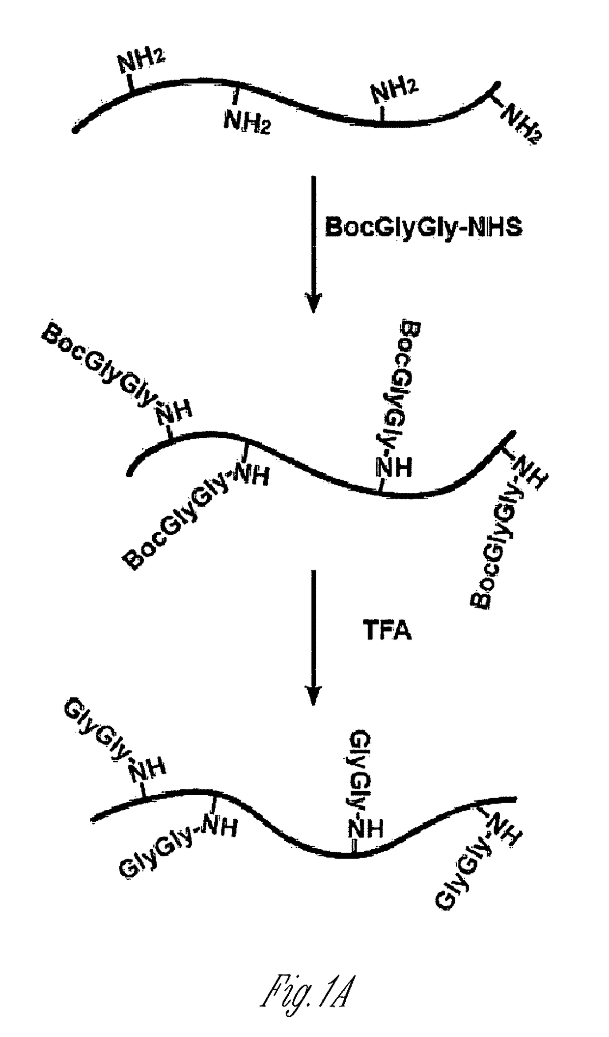
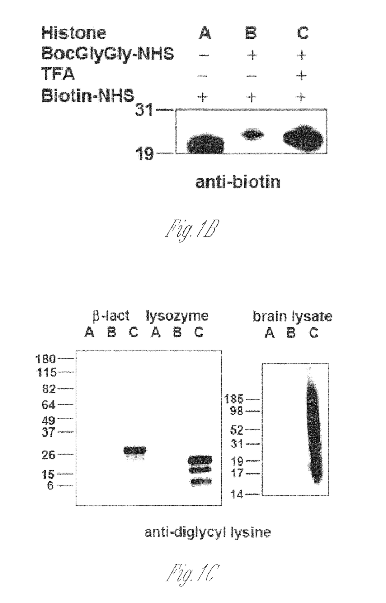
[0125]Lysine-rich histone (10 mg, sample A) from calf thymus (type III-S, Sigma) was dissolved in 100 mM NaHCO3 buffer (10 ml) at pH 10. 50 mM t-butyloxycarbonyl-Gly-Gly-N-hydroxysuccinimide (500 μl, Boc-Gly-Gly-NHS; Derrien D., et al., Glycoconj J. 1989, 6, 241-55) in DMSO was added to histone solution and the reaction was carried out at room temperature for 1 h by shaking on a plate rotator. This step was repeated three additional times and sample B was obtained. For deprotection of the Boc group, neat trifluoroacetic acid (6 ml, TFA, Sigma) was added and the solution was shaken for 2 h at room temperature. The reaction was neutralized and stopped by adding 10 M NaOH dropwise on ice (sample C). All of sample C and part of sample A and sample B were dialyzed four times against 20 mM acetic acid followed by lyop...

example 2

ng Ubiquitinated Proteins and Ubiquitination Sites

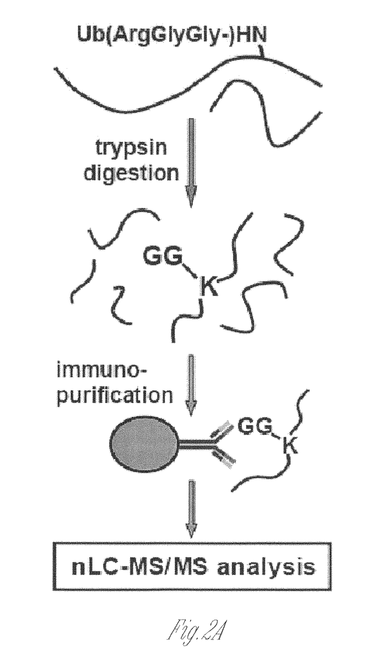
[0146]This Example illustrates novel methods for identifying ubiquitinated proteins and ubiquitination sites using an antibody that selectively binds to the diglycine remnant in peptides generated from tryptic digestion of biological samples. Using this immunoaffinity approach coupled to nano LC-MS / MS, more than 300 ubiquitinated proteins and nearly 400 ubiquitination sites were identified. Of these ubiquitinated proteins, 224 have not previously been known to be ubiquitinated. These experiments illustrate that the immunoaffinity profiling methods described herein have broad utility in characterizing the occurrence and extent of ubiquitination in diverse tissues and disease states.

[0147]To generate an antibody that recognizes peptides containing the ubiquitin remnant, a protein antigen was prepared that contained one or more diglycine adducts the terminal amine of the side chain of lysines naturally present in the protein. The scheme...

example 3

ycyl-Lysine Antibodies can be Used to Identify Ubiquitinated Ubiquitin, and the Specific Ubiquitin-Ubiquitin Linkages

[0166]Ubiquitin is often present as polyubiquitin chains, with multiple ubiquitins attached in a chain or other branched configuration on proteins. The C-terminus of ubiquitin is conjugated to other ubiquitins via one of its seven lysine residues. The antibodies described here can be used to detect the presence or abundance of specific ubiquitin chains, due to the detection of ubiquitin-specific peptides.

[0167]FIG. 12 shows a MS / MS spectrum of a fragment from ubiquitin, containing a ubiquitin remnant diglycine on K48. This peptide can be detected with either the polyclonal or monoclonal anti-diglycyl-lysine antibodies. In this case, the antibody was immobilized to protein A or protein G beads. Cellular proteins were extracted from HEK293 cells, digested by trypsin, and immunoprecipitated by GlyGly-antibody and detected on ion-trap or Q-TOF LC-MS / MS.

[0168]The detection...


PUM

PropertyMeasurementUnit
molecular weightaaaaaaaaaa
massaaaaaaaaaa
massaaaaaaaaaa

Abstract

The invention relates to particular ubiquitination epitopes, antibodies that specifically recognize and bind to ubiquitinated proteins and peptides (particularly after the ubiquitin is removed by proteolytic cleavage) and to methods of using these epitopes and antibodies.

Description

[0001]This application claims benefit of the filing date of U.S. Provisional Ser. No. 61 / 058,084, filed Jun. 2, 2008, the contents of which are specifically incorporated herein by reference.[0002]This invention was made with government support from the National Institute of Allergy and Infectious Diseases under grant number 5R21-AI068639, and from the National Cancer Institute under grant number 5T32CA062948-13. The U.S. government has certain rights in this invention.BACKGROUND OF THE INVENTION[0003]Ubiquitination has been shown to be a critical step in various cellular processes, including cell division, signal transduction, neurotransmission, and development (Bonifacino and Weissman 1998; Kirkpatrick, Denison et al. 2005), yet elucidation of the proteins targeted by ubiquitination has been difficult because of the technical challenge in recovering modified proteins. The alteration of ubiquitination pathways and patterns of ubiquitination induces many neurodegenerative diseases, s...

Claims


Application Information

Patent Timeline
Patent Type & Authority Patents(United States)
IPC IPC(8): G01N33/53C07K16/18C07K16/44G01N33/68
CPCC07K16/18G01N33/6803C07K16/44
Inventor XU, GUOQIANGJAFFREY, SAMIE R.
Owner CORNELL UNIVERSITY
Why Eureka
Try Eureka