
Medical image processing apparatus and breast image processing method thereof

a medical image processing and breast image technology, applied in image enhancement, instruments, ultrasonic/sonic/infrasonic image/data processing, etc., can solve the problems of high risk of breast cancer for women with a high density breast, and still have a high risk of false positive, so as to reduce false positives efficiently and assist density analysis of mammary glandular tissue

Active Publication Date: 2018-05-01
TAIHAO MEDICAL
View PDF15 Cites 4 Cited by
  • Summary
  • Abstract
  • Description
  • Claims
  • Application Information

AI Technical Summary

Benefits of technology

The patent text describes a medical image processing apparatus and a breast image processing method that improve the detection of mammary glandular tissue and assist in analyzing its density. The system uses texture characteristic analysis to distinguish between mammary glandular tissue and other areas in the image. This helps reduce false positive results and increases accuracy in identifying the mammary glandular tissue. Overall, the patent aims to improve the accuracy and efficiency of computer-aided detection systems for breast cancer screening.

Problems solved by technology

According to researches, it is known that compared to a low density breast, women with a high density breast has a high risk of suffering from breast cancer.
On the other hand, although a computer-aided detection (CADe) system has been used in clinical practice to automatically identify tumors, lumps, or calcifications, it still has a high risk of false positive.

Method used


Image

  • Medical image processing apparatus and breast image processing method thereof
  • Medical image processing apparatus and breast image processing method thereof
  • Medical image processing apparatus and breast image processing method thereof

Examples


Embodiment Construction

[0033]Reference will now be made in detail to the present preferred embodiments of the present disclosure, examples of which are illustrated in the accompanying drawings. Wherever possible, the same reference numbers are used in the drawings and the description to refer to the same or like parts.

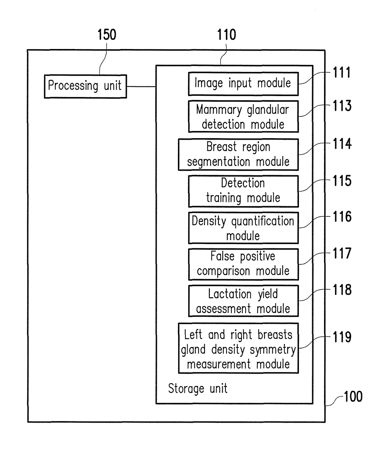
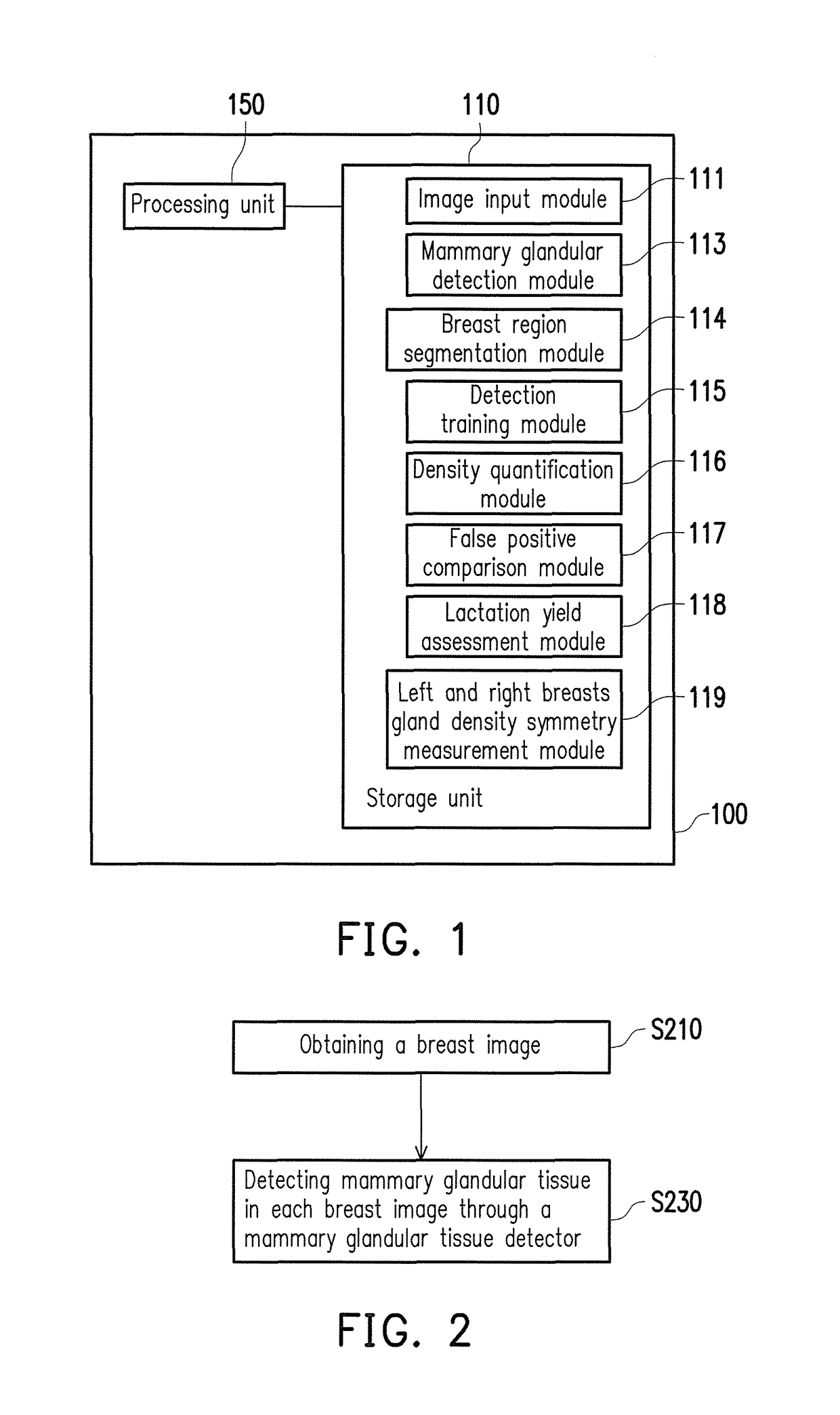
[0034]FIG. 1 is a block diagram of a medical image processing apparatus according to an embodiment of the present disclosure. Referring to FIG. 1, the medical image processing apparatus 100 at least includes (but not limited to) a storage unit 110 and a processing unit 150. The medical image processing apparatus 100 can be an electronic apparatus such as a server, a user device, a desktop computer, a notebook, a network computer, a working station, a personal digital assistant (PDA), a personal computer (PC), etc., which is not limited by the present disclosure.

[0035]The storage unit 110 may be a fixed or a movable device in any possible forms including a random access memory (RAM), a read-o...


PUM


Abstract

A medical image processing apparatus and a breast image processing method thereof are provided. The processing method at least contains but not limited to the following steps. At least one slice of breast image is obtained. Mammary glandular tissue in each breast image is detected through a mammary glandular tissue detector. The mammary glandular tissue detector is based on texture characteristic analysis. Therefore, the embodiments of the present disclosure would assist density analysis of the mammary glandular tissue and efficiently reduce false positive of computer-aided detection. In addition, based on a result of the density analysis of the mammary glandular tissue, the embodiment would further determine lactation yield and present density diagrams of mammary glandular tissue of left and right breasts. A breast region may also be separated from the breast image based on rib information according to the embodiments of the present disclosure.

Description

CROSS-REFERENCE TO RELATED APPLICATION[0001]This application claims the priority benefit of Taiwan application serial no. 105102753, filed on Jan. 28, 2016, and Taiwan application serial no. 105112377, filed on Apr. 21, 2016. The entirety of each of the above-mentioned patent applications is hereby incorporated by reference herein and made a part of this specification.BACKGROUND OF THE INVENTION[0002]1. Field of the Invention[0003]The present disclosure relates to an image processing technique, and particularly relates to a medical image processing apparatus and a breast image processing method thereof.[0004]2. Description of Related Art[0005]Mammary carcinoma is one of the most common malignant tumors in woman, and the main symptoms include breast tumor, abnormal secretions, or shape variation, etc. To early screen the abnormal breast symptoms avails treating the tumor as early as possible, so as to decrease a chance of deterioration or proliferation of cancer cells. Screening meth...

Claims


Application Information

Patent Timeline
Patent Type & Authority Patents(United States)
IPC IPC(8): G06T7/00G06T7/45G06N99/00A61B8/08A61B5/055A61B6/00
CPCG06T7/0012A61B5/055A61B6/502A61B8/0825G06T7/45G06N99/005G06T2207/30068G06T2207/10004G06T2207/20081G06F18/214G06T5/90G06N20/00G06N7/01
Inventor CHANG, RUEY-FENGCHEN, RONG-TAILAI, HSIN-HUNGCHEN, HONG-HAO
Owner TAIHAO MEDICAL
Learn More