

Apparatus and method for cone beam volume computed tomography mammography
A tomography, computerized technique used in mammography, instruments for radiological diagnosis, computed tomography scanners, etc.
- Summary
- Abstract
- Description
- Claims
- Application Information
AI Technical Summary
Problems solved by technology
Method used
Image

Examples
Embodiment Construction
[0074] Preferred and alternative embodiments of the present invention will be described in detail below with reference to the accompanying drawings, in which like reference numerals denote like parts.
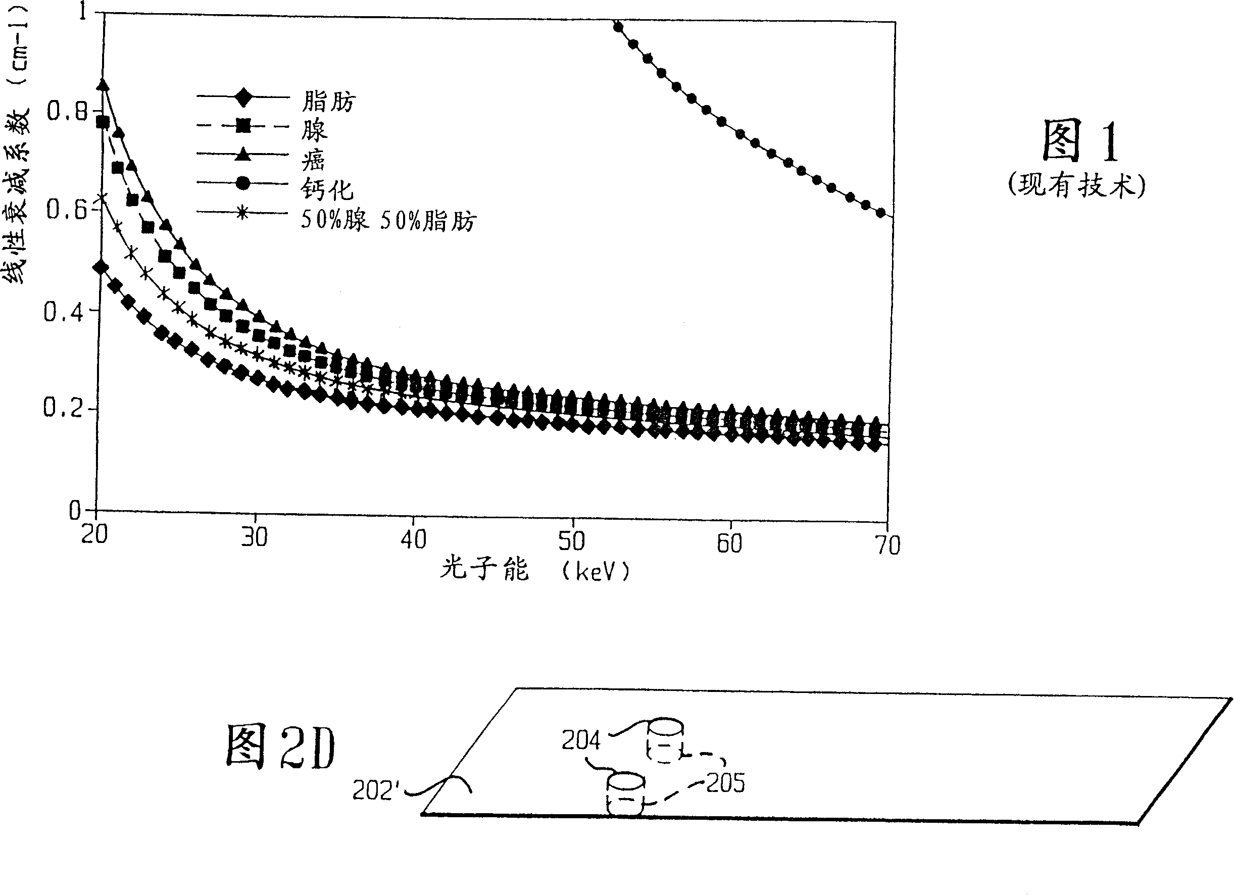
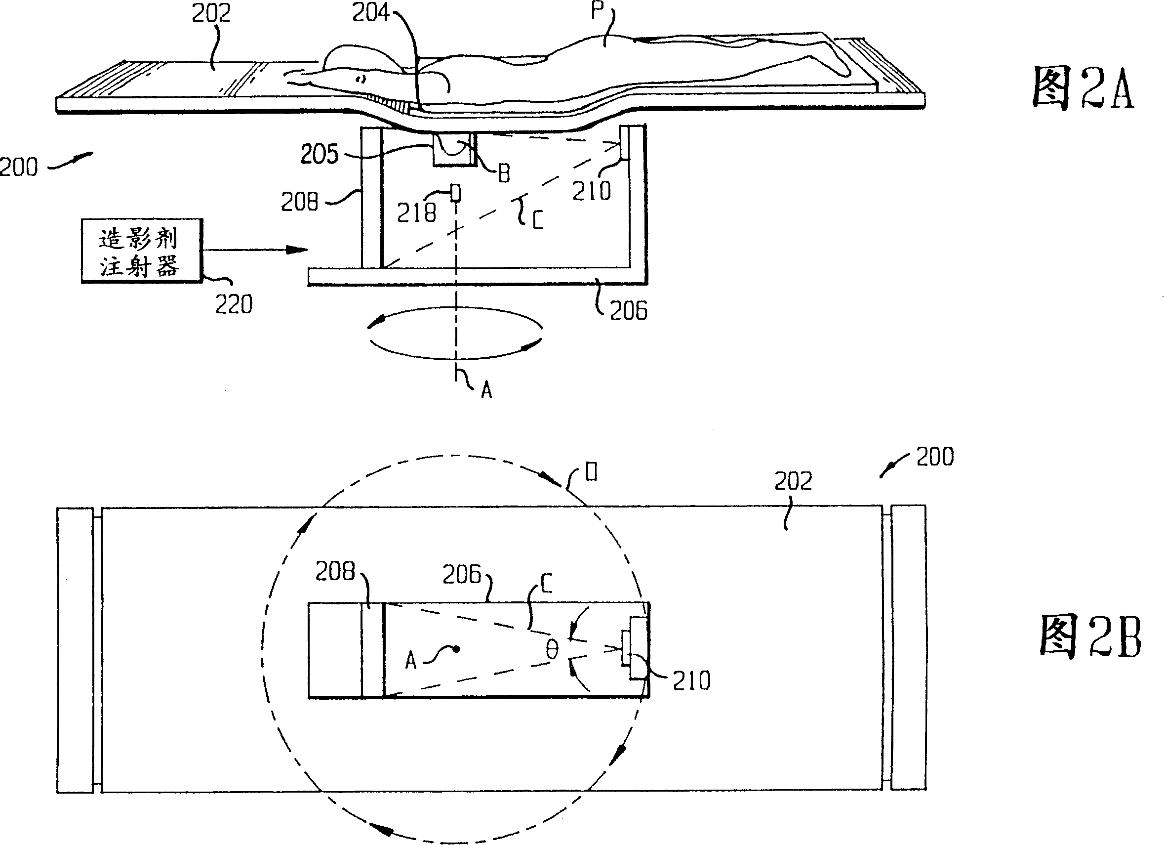
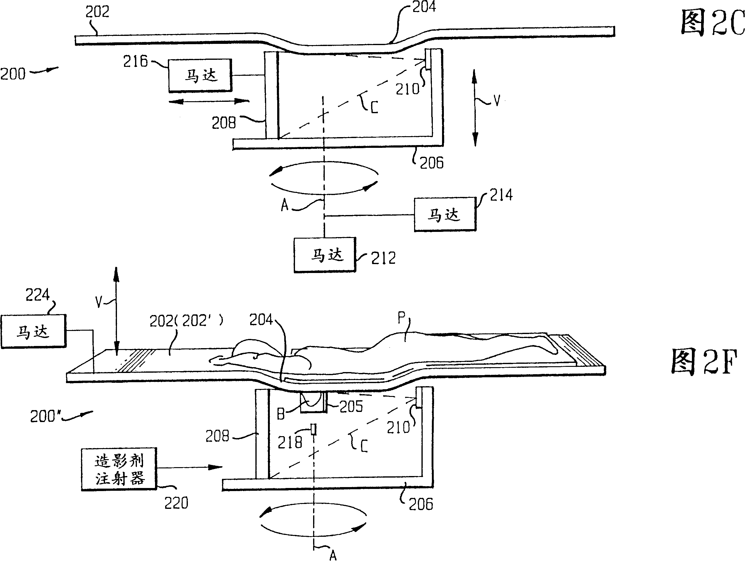
[0075] The limitations of conventional mammography will be addressed by cone beam volume CT reconstruction using flat panel detectors. With cone beam geometry and flat panel detectors, a flat panel based cone beam volumetric computed tomography mammography (CBVCTM) imaging system can be configured as shown in Figure 2A-2F, and the breast can be obtained in one fast volume scan. A three-dimensional (3D) rendering of . In contrast to conventional mammography, the tablet-based CBVC™ system is able to tomographically separate an object of interest (eg a lesion) from other objects in adjacent planes (eg other lesions or calcifications). This 3D tomographic reconstruction eliminates lesion overlap and provides a complete and correct 3D representation of breast anatomy. The CBVCTM r...
PUM
 Abstract
Description
Claims
Application Information