
Automatic division method for liver area division in multi-row spiral CT image

A CT image and multi-row helical technology, applied in computer image processing and clinical medicine, can solve problems such as easy evolution to the outside of the body cavity, failure to evolve into multiple contours, failure to obtain satisfactory results, etc., to ensure integrity Effect

Inactive Publication Date: 2010-02-10
ZHEJIANG UNIV
View PDF3 Cites 0 Cited by
  • Summary
  • Abstract
  • Description
  • Claims
  • Application Information

AI Technical Summary

Problems solved by technology

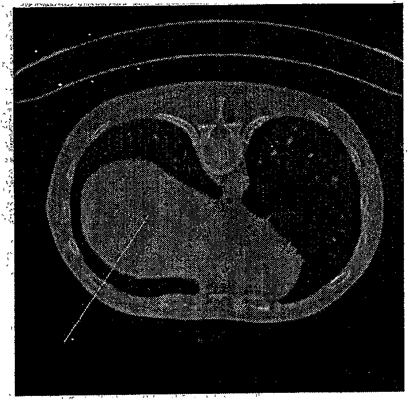
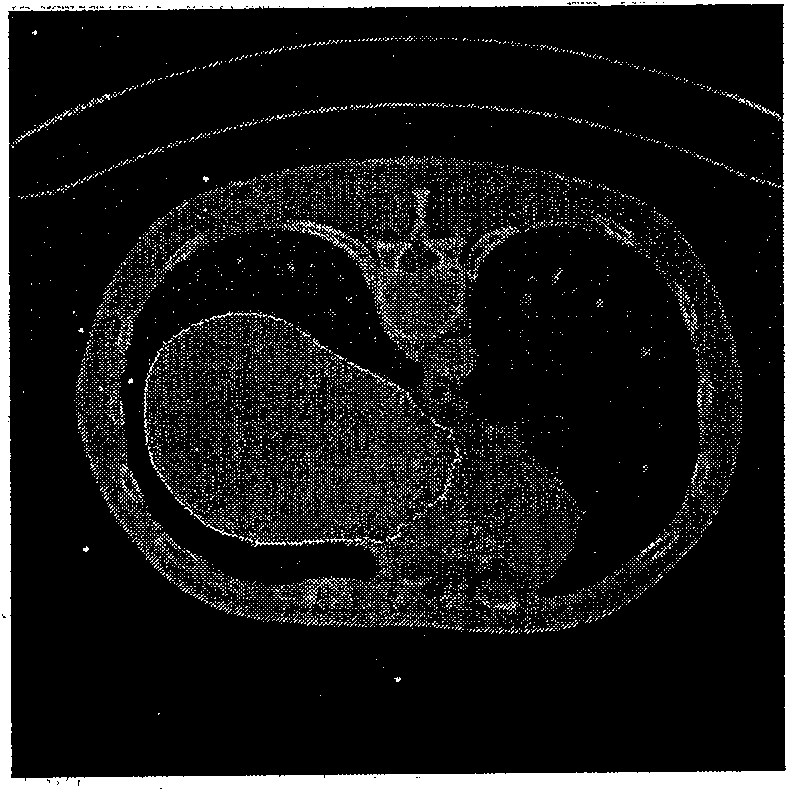
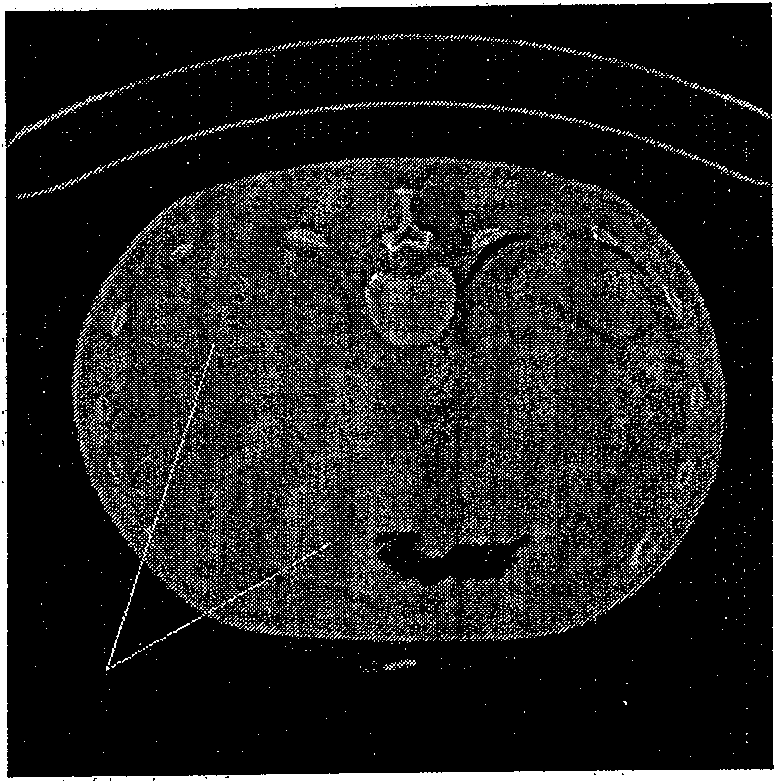
[0003] Directly applying the Fast Marching method to the liver segmentation of multi-slice spiral CT images cannot obtain satisfactory results
This is because the liver and surrounding organs such as stomach, kidney, and heart have similar gray values, so the contour will evolve to other organs without stopping; the gray value of the liver organ and the body cavity wall is also similar, resulting in the contour Easy to evolve outside the body cavity; the liver on some CT images contains two liver lobes, and the original Fast Marching method cannot evolve from one contour to multiple contours

Method used


Image

  • Automatic division method for liver area division in multi-row spiral CT image
  • Automatic division method for liver area division in multi-row spiral CT image
  • Automatic division method for liver area division in multi-row spiral CT image

Examples


Embodiment Construction

[0038] The method for automatically segmenting liver regions in multi-slice spiral CT images comprises the following steps:

[0039] 1) Select a sub-picture sequence containing the liver region in the multi-slice spiral CT image sequence, and mark the serial numbers of the first and last two pictures of the sub-sequence;

[0040] In the image sequence formed by multiple rows of spiral CT sequences, the CT image subsequence that needs to be segmented is manually determined according to medical common sense, that is, the first CT image that appears in the liver is set as the beginning image of the sequence, and the last image A CT image containing the liver is shown at the end of the sequence.

[0041] 2) Determine the region of interest ROI according to the prior knowledge of body cavity location, liver anatomical location, and liver grayscale features;

[0042] It is known from prior knowledge that the liver will not exceed the body cavity, so the body cavity area can be used...


PUM


Abstract

The invention discloses a method for automatically dividing a liver area in a multi-detector spiral CT image which helps a user to more accurately pick up the liver area in the sequence of the multi-detector spiral CT image and mark a borderline for the user to carry out further analysis and treatment on the liver area by an improved Fast Marching method and being combined with the priori knowledge of a celom location, a liver anatomical location and a liver gray characteristic. The invention realizes to intelligently assist the user to pick up the liver area from the sequence of the multi-detector spiral CT image only needing to appoint two pieces of the beginning and the tail pictures of the sub-sequence of the multi-detector spiral CT image comprising the liver without needing to mark the original profile by hand and can effectively distinguish the liver from the surrounding organs with approximate gray values to obtain an accurate liver profile, thus ensuring the user to fast obtain the liver profile and carry out further analysis and treatment. The method for automatically dividing the liver area in the multi-detector spiral CT image of the invention is suitable for assistingto decide the operation scheme of the liver surgery.

Description

technical field [0001] The invention relates to the field of computer image processing and the field of clinical medicine, in particular to a method for automatically segmenting liver regions in multi-slice spiral CT images. Background technique [0002] The Fast Marching segmentation method, the Fast Marching method proposed in J. Sethian's paper A marching level set method formonotonically advancing fronts (In Proceedings of the National Academy of Sciences, volume 93, pages 1591-1595, 1996), is through the horizontal Starting from the set method, the calculation is started from the inaccurate initial contour, and the time evolution function is applied to evolve. After reaching the termination condition, a more accurate image boundary contour is obtained. [0003] Directly applying the Fast Marching method to the liver segmentation of multi-slice spiral CT images cannot obtain satisfactory results. This is because the liver and surrounding organs such as stomach, kidney, ...

Claims


Application Information

Patent Timeline
Patent Type & Authority Patents(China)
IPC IPC(8): G06T5/00G06K9/00A61B6/03
Inventor 袁昕胡红杰王磊童海妙耿卫东
Owner ZHEJIANG UNIV
Learn More