Method for segmenting blood vessel data using serial DSA image

A digital subtraction and angiography technology, which is applied in the field of medical imaging, can solve the problems that the segmentation effect cannot detect fine blood vessels, can not achieve the denoising effect, and is unreasonable

Inactive Publication Date: 2008-02-20
HUAZHONG UNIV OF SCI & TECH
View PDF0 Cites 32 Cited by
  • Summary
  • Abstract
  • Description
  • Claims
  • Application Information

AI Technical Summary

Problems solved by technology

Since it is not judged whether there are blood vessels in the sub-block, a segmentation result will also be obtained for the background area without blood vessels, which is obviously unreasonable
Although the bimodal histogram method can be used to determine whether there are blood vessels in the sub-block to a certain extent when calculating the segmentation threshold, it requires that the number of blood vessel points in the sub-block is equal to the number of background points, which is It is very demanding and difficult to meet, and when the sub-block is small and contains a small number of pixels, it is also difficult to judge the bimodality of the histogram itself
[0006] Peng and Li (

Method used


Image

  • Method for segmenting blood vessel data using serial DSA image
  • Method for segmenting blood vessel data using serial DSA image
  • Method for segmenting blood vessel data using serial DSA image

Examples


Embodiment Construction

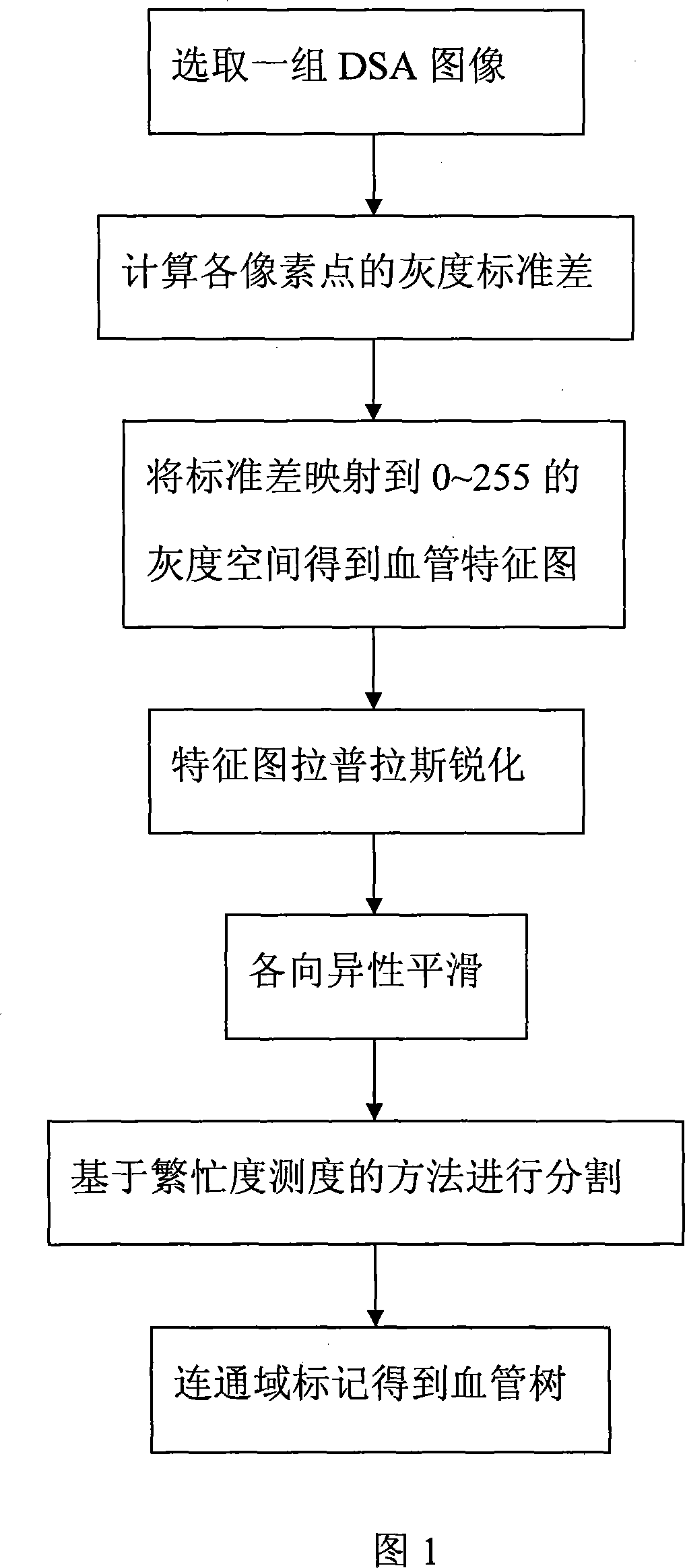
[0029] Typical embodiments of the present invention will be described below in conjunction with the accompanying drawings

[0030] Example of the present invention comprises the following steps:

[0031] (1) Select an image sequence, denoted as I(x, y, t), the image sequence I(x, y, t) includes all the images from the beginning of injection of contrast agent to the process of diffusing into blood vessels, and this one The series of images are digital subtraction angiography images registered in time;

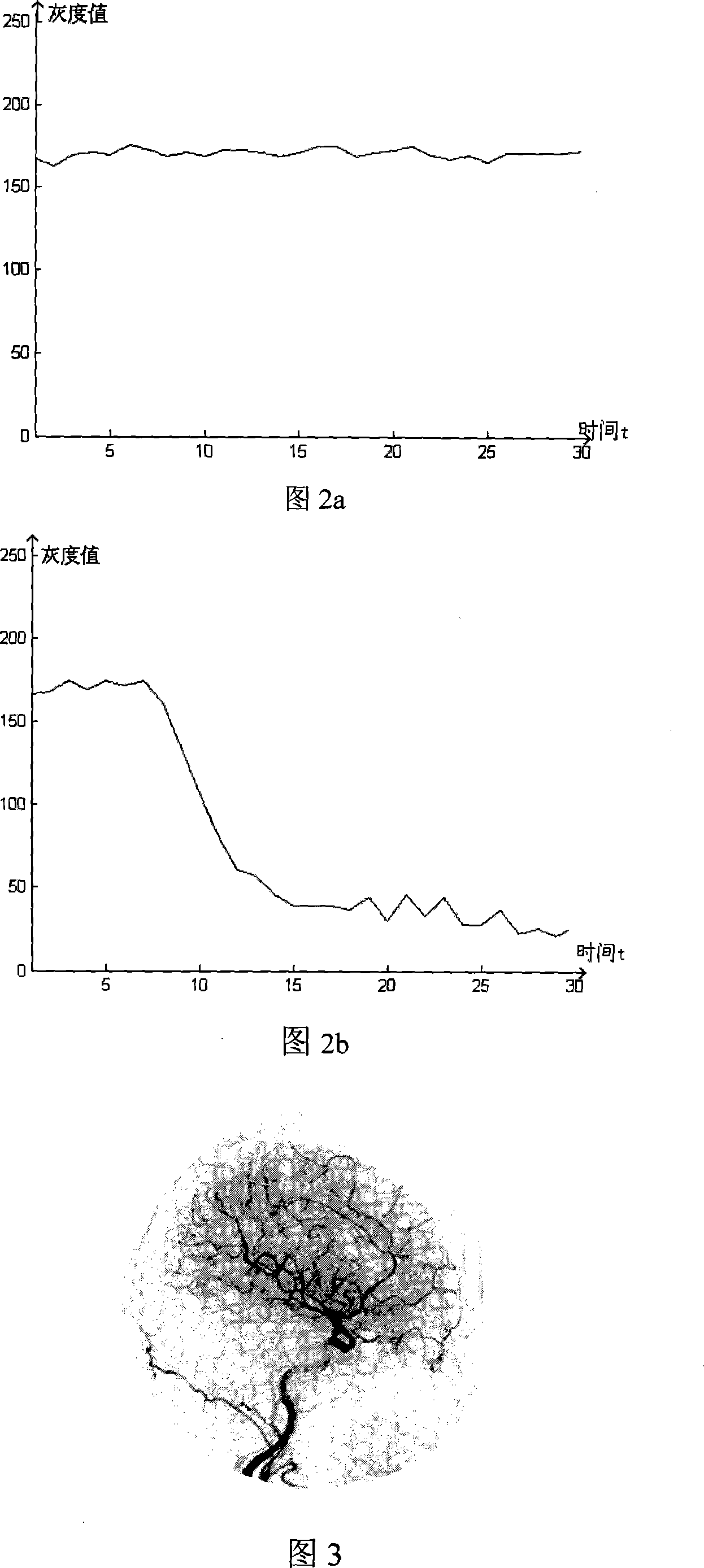
[0032] (2) Calculate the gray standard deviation value of each point on the image in the time domain, the calculation formula is: S ( x , y ) = 1 T Σ t = 1 T ( ...


PUM


Abstract

The utility model discloses a method by use of digital sequence subtraction angiographic images to segment vascular data, which makes use of standard deviation of statistics to describe the changing information of pixel gray value of each point on images along with time. By calculating the gray value standard deviation of each pixel point, 0 to 255 gray space is mapped and a feature graph describing the change of each pixel is obtained. The standard deviation of the area with bigger change of pixel gray value in time, namely, the objective vascular area, is a little bigger. The object is strengthened if reflected in the feature graph. The standard deviation of the area with smaller change of pixel gray value in time, that is the area with no blood vessel, is a little smaller. The background is weakened if reflected in the feature graph; the utility model adopts partial threshold value segmentation method on a basis of busyness measurement, carries out the binary segmentation on the feature graph, then makes use of marking technology of maximum communication domain and the connectivity of vascular tree in structure and wholly segments the vascular tree.

Description

technical field [0001] The invention belongs to the technical field of medical imaging, and in particular relates to a method for segmenting blood vessel data in digital subtraction angiography images. Background technique [0002] Digital Subtraction Angiography (DSA: Digital Subtraction Angiography) technology has been used clinically for more than 20 years, and it is an important basis for non-invasive diagnosis of cardiovascular and cerebrovascular diseases and surgical navigation for interventional therapy. A key task in DSA image processing is image segmentation, so that the physiological characteristics of blood vessels can be displayed more clearly. Subsequent operations such as structural analysis, motion analysis, and 3D visualization, as well as applied research such as image-guided surgery, tumor radiation therapy, and treatment evaluation, are all based on image segmentation. Due to the thickness of the tissues that X-rays pass through and the uneven concentrat...

Claims


Application Information

Patent Timeline
IPC IPC(8): G06T5/00A61B6/00
Inventor 桑农张天序曹治国汪春芳郭婷王国栋
Owner HUAZHONG UNIV OF SCI & TECH
Why Eureka
Try Eureka