Method and device for computerized tomoscanning vertebrae

A computer and vertebral technology, applied in the field of medical image analysis, can solve the problems of inaccurate position of intermediate vertebral slices, inconvenient clinical application of vertebral scans, small area, etc.

Active Publication Date: 2008-11-19
SIEMENS SHANGHAI MEDICAL EQUIP LTD
View PDF0 Cites 9 Cited by
  • Summary
  • Abstract
  • Description
  • Claims
  • Application Information

AI Technical Summary

Problems solved by technology

[0005] However, because the vertebra itself has the characteristics of large number, small area and inclination, especially when it is necessary to adjust or change the range of the vertebra to be scanned, it adds a lot of repetitive and time-consumi

Method used


Image

  • Method and device for computerized tomoscanning vertebrae
  • Method and device for computerized tomoscanning vertebrae
  • Method and device for computerized tomoscanning vertebrae

Examples


Embodiment Construction

[0029] In order to make the object, technical solution and advantages of the present invention more clear, the present invention will be further described in detail below in conjunction with the accompanying drawings and embodiments. It should be understood that the specific embodiments described here are only used to explain the present invention, not to limit the present invention.

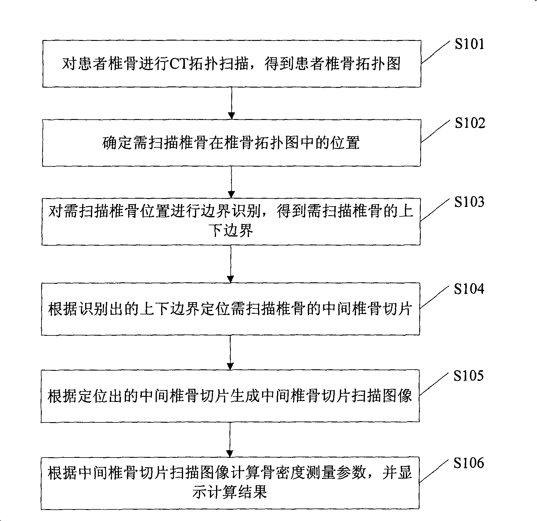
[0030] figure 1 It is a flow chart of a method for performing CT scanning on a vertebra according to an embodiment of the present invention. In this embodiment, the method of performing CT scan on the vertebrae is applied to the measurement of bone density. from figure 1 It can be seen that this embodiment specifically includes the following steps:

[0031] Step S101: performing a CT topological scan on the patient's vertebrae to obtain a topological map of the patient's vertebrae.

[0032] Step S102: Determine the position of the vertebra to be scanned in the vertebral topology map.

[003...


PUM


Abstract

The invention discloses a method for vertebrae computerized tomography (CT) scan, which comprises the steps as follows: a vertebrae topological-diagram is generated through scan; the position of the vertebrae which needs to be scanned in the vertebrae topological-diagram is determined; intermediate vertebrae slices of the vertebrae which needs to be scanned are located through graphical analysis on the vertebrae topological-diagram; the images of the intermediate vertebrae slices are generated through scan. The invention also discloses a device for the vertebrae CT scan, which comprises a topological-diagram scanning module used for generating the vertebrae topological-diagram through scan, a position determining module used for determining the position of the vertebrae which needs to be scanned in the vertebrae topological-diagram, a locating module used for locating the intermediate vertebrae slices of the vertebrae which needs to be scanned through the graphical analysis on the vertebrae topological-diagram, and a slice image scanning module used for generating the images of the intermediate vertebrae slices through scan. The method and the device can locate the intermediate vertebrae slices of the vertebrae needing to be scanned simply and conveniently during the process of vertebrae CT scan.

Description

technical field [0001] The invention relates to a medical image analysis technology, in particular to a method and a device for performing computed tomography (CT, Computed Tomography) on a vertebra. Background technique [0002] At present, the measurement of bone density usually adopts non-invasive bone density measurement method, which usually consists of two parts. The first part is the scanning process, including CT topographic scan (also known as CT positioning image scan, Topogram), middle vertebra Slice location or slice detection and CT scan of intermediate vertebral slices, etc.; the second part is the evaluation process, including result display and evaluation parameter comparison, etc. [0003] The CT scan mentioned here refers to scanning a layer of a certain thickness of a certain part of the human body with an X-ray beam, and the detector receives the X-ray passing through this layer, and the measured signal is converted by analog-to-digital conversion (ADC) ...

Claims


Application Information

Patent Timeline
IPC IPC(8): A61B6/03G06F5/00
Inventor 张颖
Owner SIEMENS SHANGHAI MEDICAL EQUIP LTD
Why Eureka
Try Eureka