Liver tumor segmentation method and device based on CT (Computed Tomography) image

A technology for CT images and liver tumors, applied in the field of medical image processing, to achieve good separability, accurate and robust segmentation, and optimize tumor segmentation results

Active Publication Date: 2016-05-11
SHENZHEN INST OF ADVANCED TECH CHINESE ACAD OF SCI +1
View PDF3 Cites 92 Cited by
  • Summary
  • Abstract
  • Description
  • Claims
  • Application Information

AI Technical Summary

Problems solved by technology

[0004] The technical problem to be solved in the present invention is to overcome th

Method used


Image

  • Liver tumor segmentation method and device based on CT (Computed Tomography) image
  • Liver tumor segmentation method and device based on CT (Computed Tomography) image
  • Liver tumor segmentation method and device based on CT (Computed Tomography) image

Examples


Embodiment Construction

[0018] The following will clearly and completely describe the technical solutions in the embodiments of the present invention with reference to the accompanying drawings in the embodiments of the present invention. Obviously, the described embodiments are only some, not all, embodiments of the present invention. Based on the embodiments of the present invention, all other embodiments obtained by persons of ordinary skill in the art without making creative efforts belong to the protection scope of the present invention.

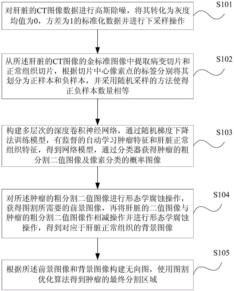
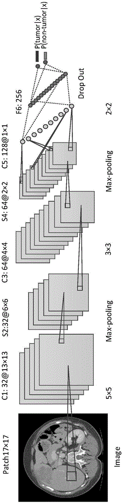
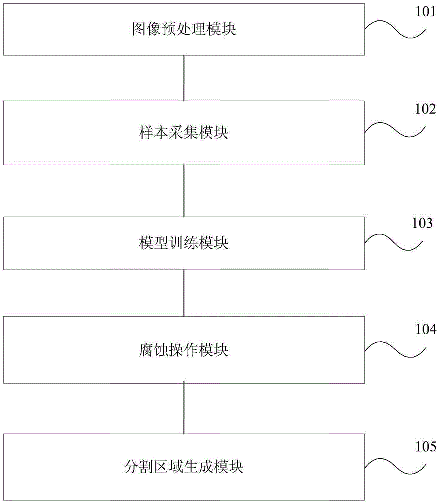
[0019] The invention discloses a fully automatic liver tumor segmentation method based on CT images, the main process of which is: performing denoising, standardization, and down-sampling preprocessing operations on a series of original CT liver images; training images and test images Separately extract the positive and negative samples required for training and the samples required to predict the label for testing; train the deep convolutional neural network m...


PUM


Abstract

The invention provides a liver tumor segmentation method and device based on a CT (Computed Tomography) image. The method comprises the following steps: performing Gaussian denoising on CT image data of a liver, converting the denoised CT image data into standardized data of which a gray average is 0 and a variance is 1, and performing down-sampling operation; extracting a lesion slice and a normal tissue slice from a gold standard image of the CT image of the liver, and classifying the lesion slice and the normal tissue slice into a positive sample and a negative sample; constructing a multi-level depth convolutional neural network, training a model through a stochastic gradient descent to obtain a network model, and acquiring a coarse segmentation binary image of a tumor and a pixel-classification probability image through a classifier; performing morphological erosion operation on the coarse segmentation binary image of the tumor to obtain a foreground image needed by graph cut, performing subtraction operation on the binary image of a liver and the coarse segmentation binary image of the tumor, and performing the morphological erosion operation to obtain a background image corresponding to normal tissues of the liver; and constructing an undirected graph, and obtaining a finial segmentation region of the tumor through a graph cut optimization algorithm.

Description

technical field [0001] The invention belongs to the field of medical image processing, in particular to a CT image-based liver tumor segmentation method and device. Background technique [0002] The liver is an important and complex functional organ to maintain the life activities of the human body. There are many types of liver lesions, and the incidence rate is high. Computed Tomography (CT) images have become one of the important routine means in clinical diagnosis and an important means of examination for liver diseases. Currently, the treatments for liver tumors mainly include tumor resection, interventional therapy, and radiotherapy, among which tumor resection is the most effective treatment. These treatment methods all require accurate knowledge of the number, location, size, and shape of the tumor before surgery, which is helpful for the formulation of a treatment plan for liver tumors. , size, shape, gray scale and texture are different, it is difficult to develo...

Claims


Application Information

Patent Timeline
IPC IPC(8): G06T7/00
CPCG06T2207/10081G06T2207/30056
Inventor 贾富仓李雯贺宝春胡庆茂方驰华范应方
Owner SHENZHEN INST OF ADVANCED TECH CHINESE ACAD OF SCI
Why Eureka
Try Eureka