Apparatus for simultaneous lossless detection of cell and extracellular matrix component

A non-destructive detection and cell technology, applied in the direction of material excitation analysis, fluorescence/phosphorescence, etc., can solve the problems of inaccurate staining time, loss of accuracy of inspection results, inappropriate specimen handling, etc.

Inactive Publication Date: 2009-08-12
FUJIAN NORMAL UNIV
View PDF0 Cites 27 Cited by
  • Summary
  • Abstract
  • Description
  • Claims
  • Application Information

AI Technical Summary

Problems solved by technology

In addition, inappropriate handling of specimens and improper

Method used


Image

  • Apparatus for simultaneous lossless detection of cell and extracellular matrix component

Examples


Embodiment Construction

[0007] Embodiments of the present invention will be further described below in conjunction with the accompanying drawings, so as to make the present invention more comprehensible.

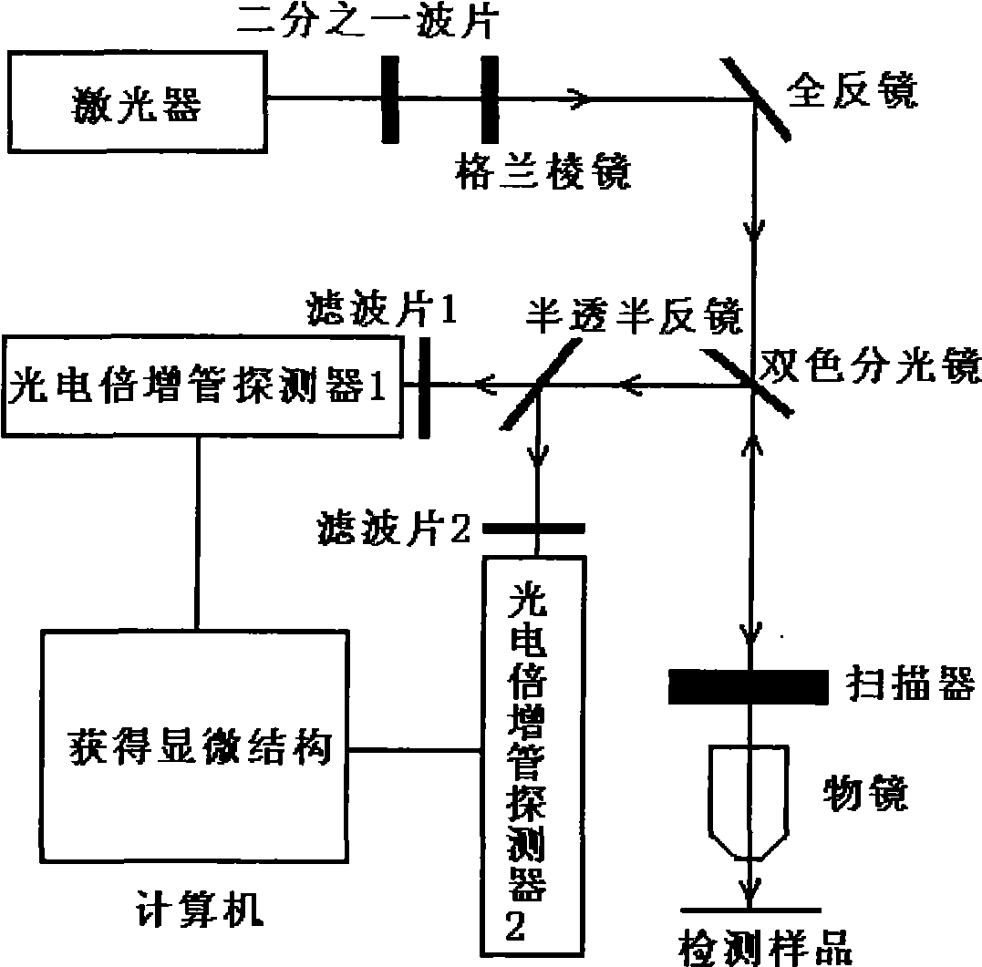
[0008] Such as figure 1 As shown, the light emitted by the laser is incident on the scanning mirror group of the scanner through a two-color beam splitter, and the beam passing through the scanning mirror group is focused on the detection sample by the objective lens; the two-photon excitation fluorescence and the second harmonic generated by the laser and the detection sample The wave signal is collected by the same objective lens in reverse, and then incident on a half-transparent mirror by the aforementioned scanning mirror group and dichroic beam splitter, and the emitted signal light composed of two-photon excited fluorescence and the second harmonic signal is divided into two paths, One path converts the second harmonic signal of collagen fibers into electrical signal A through filter 1 and p...


PUM


Abstract

The invention relates to a device for performing synchronous nondestructive detection on intercellular and extracellular matrix components, which is characterized in that light emitted by a laser is incident onto a scanning mirror group of a scanner through a dichroic beam splitter, and light beams which pass through the scanning mirror group are focused on a detected sample by an objective lens; two-photon excited fluorescence and a secondary harmonic signal produced by the laser and the detected sample reversely passes through the same objective lens to be collected, then are incident onto a half transparent and half reflecting mirror by the scanning mirror group and the dichroic beam splitter; and emission signal light consisting of the two-photon excited fluorescence and the secondary harmonic signal is divided into two paths, the two paths are converted into electric signals through a filter plate and a photomuitplier and are connected to an input end of a computer, and finally the computer synchronously displays micro-structures of cells, elastic fibers and collagenous fibers. The device can perform high-resolution, high-contrast, nondestructive, quick and real-time detection on the microstructures of the cells, the elastic fibers and the collagenous fibers at molecular level.

Description

technical field [0001] The present invention relates to a device for simultaneous non-destructive probing of cells and components of the extracellular matrix. technical background [0002] At present, the pathology department of clinical medicine observes and detects the microstructure of cells, collagen fibers and elastic fibers, the main components of extracellular matrix, every day. The detection methods of clinical medicine are usually traditional histopathological methods. Pathological tissue sections for histopathological analysis need to be obtained through a series of processes such as fixation, paraffin embedding, dewaxing, and staining. These processes have not only destroyed some relevant and useful material real forms of the tissue, but also require a relatively long time. Long, the time spent on each step is calculated in hours or even days. In addition, inappropriate handling of specimens and inaccurate staining time may lead to loss of accuracy of test resul...

Claims


Application Information

Patent Timeline
IPC IPC(8): G01N21/64
Inventor 陈建新卓双木谢树森姜兴山刘嫩容卢可成
Owner FUJIAN NORMAL UNIV
Why Eureka
Try Eureka