

Biological implantation material and method for preparing same
An implant material and biological technology, applied in the field of biological implant materials and their preparation, can solve the problem of difficult to remove cells and the like
- Summary
- Abstract
- Description
- Claims
- Application Information
AI Technical Summary
Problems solved by technology
Method used
Image

Examples
Embodiment 1
[0048] Example 1 : Preparation of endothelial implant material
[0049] Bovine afterbirth samples collected from bovine placenta were cryopreserved in sterile saline and shipped to the laboratory. will be 500cm 2 The collected samples were treated with 1 liter of 95% ethanol overnight in a cold room, which removes the fat from the bovine afterbirth samples. Wash the sample with 1L of pure water for three times, 10 minutes each time; use a spatula to remove the matrix layer in the sample. The above sample was treated with 1 liter of 70% ethanol at low temperature to inactivate the virus, and then 1 liter of EDTA / NaCl solution (pH 11), which contained 0.2% EDTA and 0.9% NaCl, was added and stirred at 150rpm for 1 hour To remove soluble alkaline impurities (step (i)). Then, an enzymatic digestion reaction was performed with a solution containing 0.05% trypsin, 0.02% EDTA and 0.9% sodium chloride (pH 7.4), which was stirred at 37°C for 1 hour (step (ii)). The obtained cell c...
Embodiment 2
[0052] Example 2 : Content of fat and modified collagen
[0053] Determination of the effect of the following three methods, that is, the method of Example 1, according to the method (condition A) of Ginger A. Abraham (US Patent No. 2006 / 0024380) and according to the method of Tooru Yui et al. (US Patent No. 5,876,451) (Condition B). The method established according to conditions A and B is described in detail below.
[0054] In condition A, the afterbirth matrix derived from bovine placenta was removed. Add 1 liter of 0.1M EDTA / 10mM NaOH solution per 100cm2 sample, stir at 200rpm for 18 hours, then add 1 liter of 1M HCl / 10mM NaOH solution, and stir at 200rpm for 8 hours. The obtained sample was treated with 1 L of 1M NaCl / 10 mM phosphate buffer solution (PBS), stirred for 18 hours, 1 L of 10 mM PBS was added thereto and stirred for 2 hours, and further stirred in sterile purified water at 200 rpm for 1 hour.
[0055] In condition B, the afterbirth matrix derived from bov...
Embodiment 3
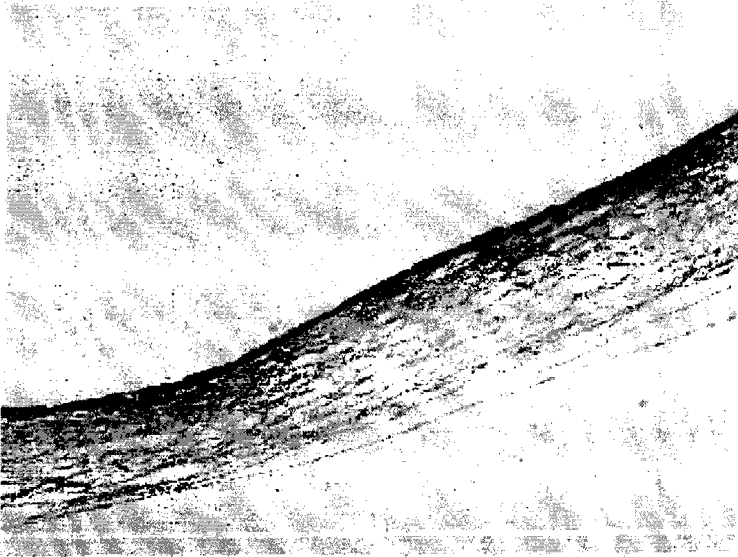
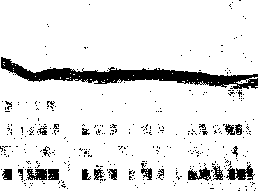
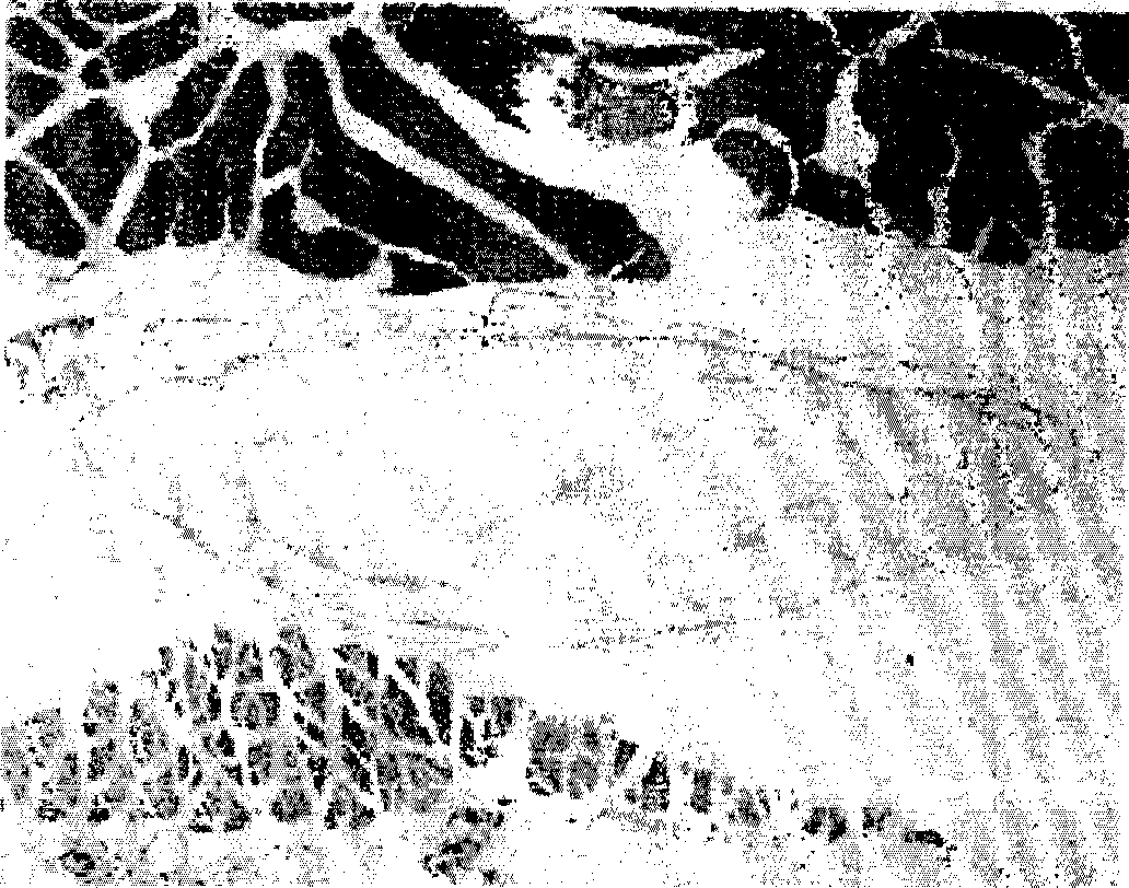
[0066] Example 3 : Biocontainment Test of Subcutaneous Implantation in Guinea Pigs
[0067] The degree of inflammation of inflammatory cells and calcification in vivo were detected by subcutaneous injection in guinea pigs. The operation steps are to subcutaneously implant the endothelial implant material and Surgisis prepared in Example 1 respectively in different guinea pigs. TM (Cook Inc. USA), and then compare the above-mentioned guinea pig tissues of different treatments. After 2 and 4 weeks, guinea pig tissues from different treatments were fixed with formalin, washed or coated with parapins. The above-treated tissues were sectioned to a thickness of 5 μm, stained with hematoxylin and eosin (H&E), and observed by an optical microscope. After 2 weeks and 4 weeks, the micrographs of the H&E staining of the tissues of subcutaneous implantation of the aftercoat implant material prepared in embodiment 1 are respectively as follows: Figure 2a and 2b indicated, after 2 and...
PUM
 Abstract
Description
Claims
Application Information