Integrated method for enriching and detecting rare cell in biological fluid sample

A technology of rare cells and biological fluids, applied in biochemical equipment and methods, animal cells, biological testing, etc., can solve the problems of restricting the development of circulating tumor cells and circulating endothelial cells, and achieve the effect of promoting popular application and low cost

Active Publication Date: 2009-11-25
CYTTEL BIOSCI BEIJING
View PDF3 Cites 64 Cited by
  • Summary
  • Abstract
  • Description
  • Claims
  • Application Information

AI Technical Summary

Problems solved by technology

However, its unavoidable high non-specific staining signal, expensive fluorescence microscope, and necessary but inconvenient working environment (such as dark room) greatly limit the detection of circulating tumor cells and circulating endothelial cells based on immunofluorescence.

Method used


Image

  • Integrated method for enriching and detecting rare cell in biological fluid sample
  • Integrated method for enriching and detecting rare cell in biological fluid sample

Examples


Embodiment 1

[0064] Example 1. Enrichment of circulating tumor cells from the peripheral blood of breast cancer patients

[0065] 5ml of human peripheral blood was collected in blood collection tubes (BD, New Jersey, USA) containing ethylenediaminetetraacetic acid (EDTA) anticoagulant. After centrifuging the blood sample (700xg, 10 minutes), the supernatant can be aspirated using a pipette or automatic pipetting device to remove plasma proteins. The pellet obtained after centrifugation was resuspended in 30 ml of erythrocyte lysate (BD Pharmingen, California, USA) and incubated for 20 minutes. Specimens were centrifuged (700xg, 10 minutes) to separate the lysed erythrocyte fragments in the supernatant. After removing the supernatant, resuspend the pellet (ie, deposited cells) in 5 mL of phosphate buffer (pH 7.4). After adding 0.5 ml of magnetic beads coated with monoclonal antibody against leukocyte surface antigens such as CD45 (Invitrogen, California, USA) therein, incubate at room tem...

Embodiment 2

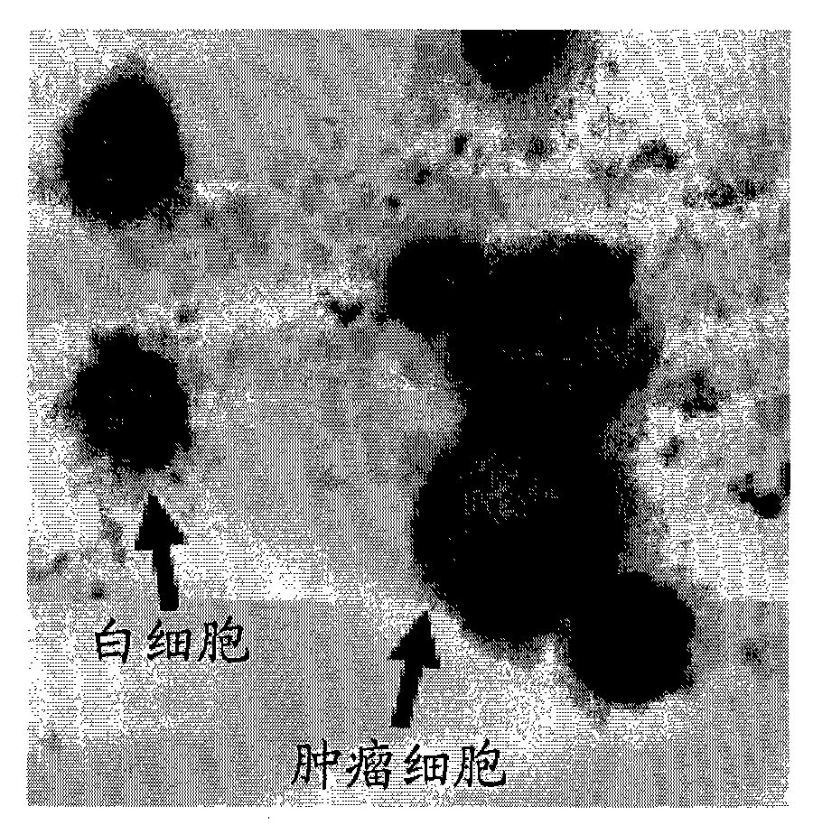
[0067] Example 2. Trichrome staining of circulating tumor cells enriched from peripheral blood of breast cancer patients

[0068] The enriched circulating tumor cells were placed on a glass slide, fixed with 2% paraformaldehyde (paraformaldehyde) prepared from phosphate buffered saline for 2 hours at room temperature, and washed 3 times with phosphate buffered saline. Cells were treated with biotin (Pierce, Illinois, USA)-labeled anti-keratin 8+18+19 monoclonal antibody (Abcam, UK, 1 μg / ml) and rhodamine (Pierce, Illinois, USA)-labeled anti-CD45 monoclonal antibody Antibody (Abcam, UK, 1 μg / ml) mixture (diluted from phosphate buffer) was incubated at room temperature for 30 minutes. After the slides were washed three times with phosphate buffered saline, they were labeled with alkaline phosphatase-labeled anti-biotin monoclonal antibody (Sigma, Missouri, USA, 1 μg / ml) and peroxidase (Pierce, Illinois, USA) Anti-rhodamine monoclonal antibody (Abcam, UK, 1 μg / ml) mixture (dilut...

Embodiment 3

[0070] Example 3. Detection of circulating tumor cells by chromosome fluorescence in situ hybridization

[0071] The enriched tumor cells were placed on slides as specimens. After the stained specimen was treated with 20 mg / ml RNase for 1 hour, the slide was rinsed with SSC buffer. The specimens were dehydrated with absolute ethanol for 10 minutes, then heated to 70°C for 5 minutes to denature. The specimen was then dehydrated with absolute ethanol for 10 minutes, and incubated overnight at 45°C for hybridization with the probe. After the specimens were washed with SSC buffer, they were observed with a fluorescence microscope. The specimen may be enriched tumor cells stained by the method in Example 2, and the purpose of performing chromosomal fluorescence in situ hybridization is to further confirm the authenticity of detecting tumor cells based on immunohistochemical trichrome staining. In order to facilitate rapid diagnosis, specimens can also be directly subjected to ch...


PUM

PropertyMeasurementUnit
diameteraaaaaaaaaa

Abstract

The invention relates to an integrated method for enriching and detecting a rare cell in a biological fluid sample. The enriching method comprises the following steps: (a) removing plasma protein by centrifugation; (b) optionally adding hematid lysis solution to carry out hematid lysis so as to remove hematid; (c) adding an immune microsphere or immune adsorbent to incubate; and (d) carrying out density centrifugation based on a specific cell separation medium so as to separate the a cycling rare cell, the residual cell after the hematid is removed and leucocyte combined on the immune microsphere. The method for detecting the enriched rare cells comprises the steps of combining pigmentation based on immune histochemistry or bi-color, tri-color or multicolor pigmentation based on the immunehistochemistry with immune fluorescence and observing and identifying by a fluorescent or common optical microscope or a scanner based on a microscope principle. The novel and unique method for enriching and detecting a rare cell in a biological fluid sample is proved to have low cost and can rapidly, effectively and highly specifically enrich and quantitatively detect the rare cell in blood.

Description

technical field [0001] The present invention mainly relates to an integrated method for enrichment and detection of rare cells from biological fluid samples. Background technique [0002] Since the U.S. Food and Drug Administration approved the technology of directly capturing circulating tumor cells in human peripheral blood from Immunicon / Veridex (Philadelphia, USA) in 2004, the acquisition and detection of circulating tumor cells, circulating endothelial cells, and tumor stem cells , and the important scientific research and clinical significance of certain immune cells have been widely reported (Cristofanilli et al, 2004 New Eng J.Med.351:781; Braun and Marth, 2004 New Eng.J.Med.351:824 ). [0003] However, this method of using antibody-coupled magnetic beads to directly capture circulating tumor cells has a well-known disadvantage (Mocellin et al, 2006 Trends in Molecular Medicine12:130): due to the heterogeneity of expression of tumor cell surface markers, many tumor ...

Claims


Application Information

Patent Timeline
Patent Type & Authority Applications(China)
IPC IPC(8): G01N1/40G01N1/34G01N33/48C12Q1/00C12Q1/24
CPCG01N33/6893G01N33/5011G01N33/5005C12N5/0693G01N33/6803G01N33/574
Inventor 林平
Owner CYTTEL BIOSCI BEIJING
Why Eureka
Try Eureka