Anti-segmentation method for medical image

A medical image and three-dimensional image technology, applied in image analysis, image data processing, 3D image processing, etc., can solve the problems of only viewing, high hardware parameters, slow execution speed, etc., and achieve the effect of visualization

Inactive Publication Date: 2010-05-19
SOUTH CHINA NORMAL UNIVERSITY
View PDF0 Cites 14 Cited by
  • Summary
  • Abstract
  • Description
  • Claims
  • Application Information

AI Technical Summary

Problems solved by technology

Its disadvantages are: 1) Process pause and result modification operations cannot be performed during the segmentation process, only viewing and rotation
2) The selection of the seed point is carried out on the two-dimensional image, and it cannot be accurately judged whether it belongs to the point of the target segmentation object
3) The hardware parameters required by the technology are relatively high
4) The execution speed of the method is slow, and it takes a long time to get the result
Its disadvantage is: the main disadvantage of the method used by the system is that the user still cannot operate and transform the data during the extraction and display process
Moreover, the surface model extraction algorithm used is relatively complex and the running speed is relatively slow, requiring users to wait for a long time
And there are also many parameters that need to be set based on experience

Method used


Image

  • Anti-segmentation method for medical image
  • Anti-segmentation method for medical image

Examples


Embodiment

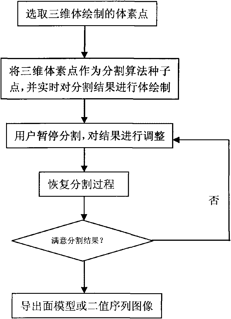
[0043] The principle block diagram of the present invention is as figure 1 Shown, the medical image anti-segmentation method of the present invention, it comprises the following steps:

[0044] 1) using a two-dimensional input device to select corresponding three-dimensional voxel points from the three-dimensional three-dimensional organ rendered by three-dimensional volume;

[0045] 2) Use the 3D voxel point as the seed point of the segmentation algorithm, and perform volume rendering on the segmentation result in real time; and display the volume rendering of the segmentation result in the 3D image space in real time during the algorithm segmentation process;

[0046] 3) During the volume rendering display process of the segmentation result, when the user observes that the segmentation result does not meet his requirements, he can immediately suspend the segmentation, adjust the unreasonable part, and then resume the segmentation process;

[0047] 4) The user can adjust the...


PUM


Abstract

The invention relates to an anti-segmentation method for a medical image, which comprises the following steps that: 1) two-dimensional input equipment is used for selecting a corresponding three-dimensional voxel point from a three-dimensional organ by three-dimensional volume rendering technology; 2) the three-dimensional voxel point is used as a seed point of a segmentation arithmetic, a segmentation result is subjected to volume rendering, and the segmentation result is displayed in the space of a three-dimensional image during the segmentation arithmetic in real time by volume rendering; 3) in the displaying process of the segmentation result by volume rendering, a user can pause the segmentation step at once when observing that the segmentation result does not accord with the requirement of the user, adjust the unreasonable part and restore the segmentation process; 4) the user can adjust the segmentation result in real time in the whole segmentation process until the user is satisfied with the segmentation result; and 5) when the user is satisfied with the current segmentation result, the user can save and export the current segmentation result at once into a binary sequenceimage, and also can perform surface rendering on the current segmentation result at once, export a surface model of the result and prepare for the subsequent virtual operation. Through the method, the user can basically obtain a precise abdominal organ object by performing once segmentation process so as to provide exact data for the subsequent virtual operation.

Description

technical field [0001] The invention is a medical image anti-segmentation method, which belongs to the innovative technology of the medical image anti-segmentation method. Background technique [0002] Image segmentation is one of the key steps in 3D reconstruction of CT tomographic data. Separate different areas in the image that have special meanings that do not cross each other, so that each area meets the consistency of a specific area. Image segmentation has special significance in medical applications. It is a key step in medical image processing and a comprehensive research topic spanning medicine and computer science. Medical image segmentation is the basis for lesion region extraction, specific tissue measurement, and 3D reconstruction. Due to factors such as imaging principles, local volume effects, and blurred tissue edges, the segmentation of 3D medical data fields has always been a challenging topic. So far, there is still no general theory for medical data f...

Claims


Application Information

Patent Timeline
Patent Type & Authority Applications(China)
IPC IPC(8): G06T7/00G06T15/00
Inventor 杨璐彭丰平潘家辉
Owner SOUTH CHINA NORMAL UNIVERSITY
Why Eureka
Try Eureka