
Related prediction model-based method for detecting structural deformation in magnetic resonance image
A technology of magnetic resonance images and nuclear magnetic resonance images, which is applied in the field of medical image processing of magnetic resonance images, can solve the problems of objectivity and lack of repeatability, and achieve the effect of precise geometric structure
- Summary
- Abstract
- Description
- Claims
- Application Information
AI Technical Summary
Problems solved by technology
Method used
Image

Examples
Embodiment Construction
[0034] Now in conjunction with embodiment, accompanying drawing, the present invention will be further described:
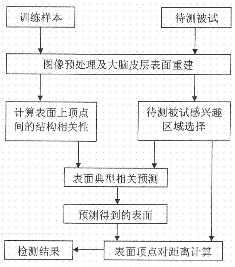
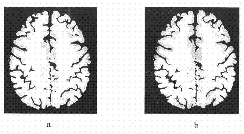
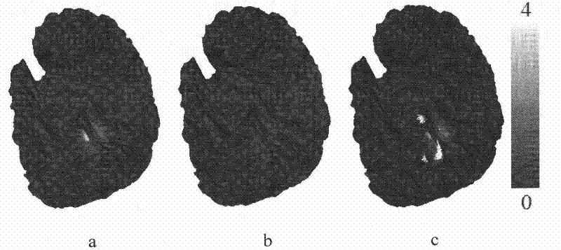
[0035]Steps in this embodiment: 1) For a group of normal subjects, reconstruct the surface of the cerebral cortex according to their magnetic resonance structural images, and use the elastic deformation registration algorithm to register all the cortical surfaces to the standard template space to construct a set of training samples , and then use the training samples to calculate the structural correlation matrix between vertices on the surface of the cerebral cortex; 2) For the sample to be tested, reconstruct the surface of the cerebral cortex according to its magnetic resonance structural image; 3) On the surface of the cerebral cortex of the subject to be tested Select the area to be detected, and according to the structural correlation matrix calculated in step 1), use the typical correlation prediction model to predict the expected coordinate value of the ve...
PUM
 Abstract
Description
Claims
Application Information