Ultrasound imaging apparatus, image processing apparatus and image processing method

A photographic device and ultrasonic technology, applied in image data processing, ultrasonic/sonic/infrasonic diagnosis, sound wave diagnosis, etc., can solve problems such as not knowing the position of the detector and difficult to recognize the direction of object movement

Active Publication Date: 2010-09-01
TOSHIBA MEDICAL SYST CORP
View PDF2 Cites 9 Cited by
  • Summary
  • Abstract
  • Description
  • Claims
  • Application Information

AI Technical Summary

Problems solved by technology

[0006] However, when the 3D image is rotated and the color Doppler display is performed on any section, the display is still based on the position of the detector, but since the position of the detector is not known, it is difficult to recognize the direction of motion of the object

Method used


Image

  • Ultrasound imaging apparatus, image processing apparatus and image processing method
  • Ultrasound imaging apparatus, image processing apparatus and image processing method
  • Ultrasound imaging apparatus, image processing apparatus and image processing method

Examples


Embodiment Construction

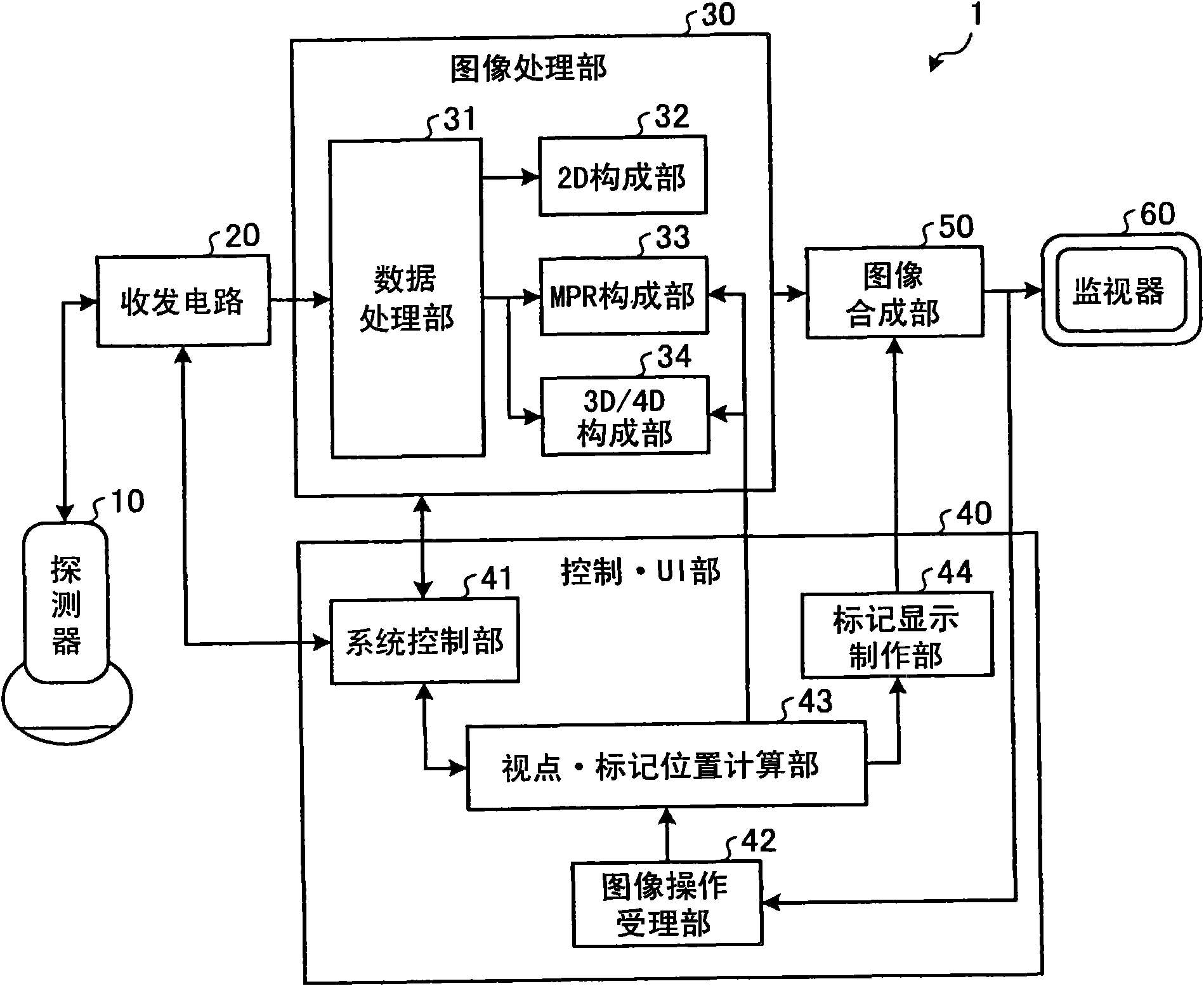
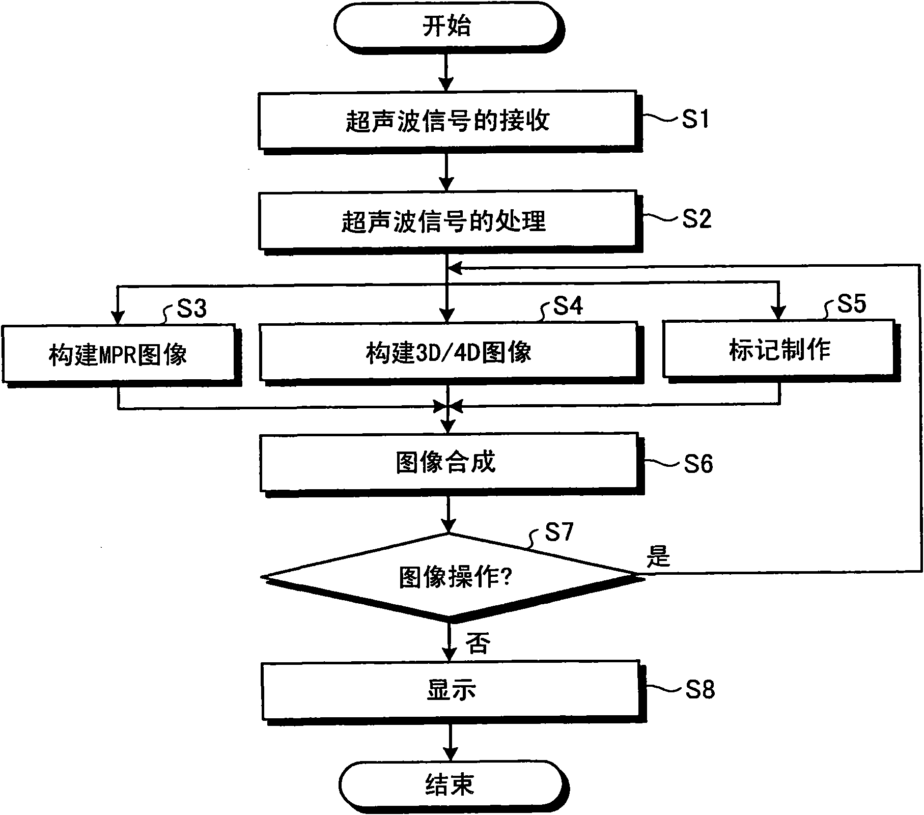
[0015] Hereinafter, preferred embodiments of the ultrasonic imaging apparatus, image processing apparatus, and image processing method of the present invention will be described in detail with reference to the accompanying drawings.

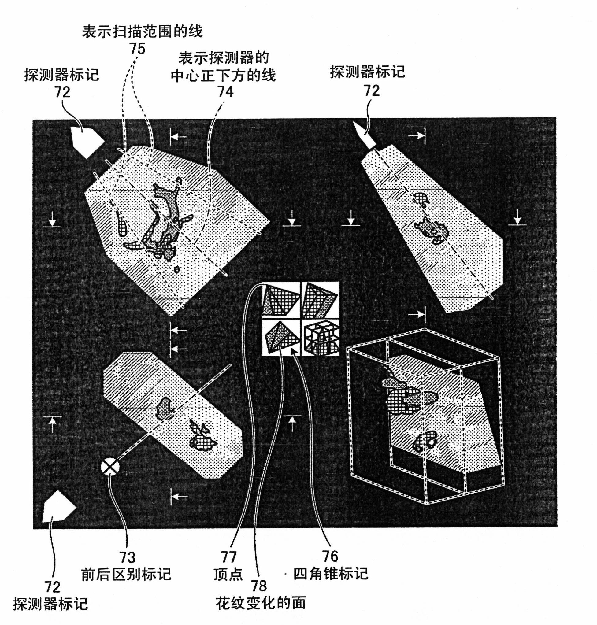
[0016] First, an MPR image and a 3D image displayed by the ultrasonic diagnostic apparatus of this embodiment will be described. figure 1 It is a diagram showing an example of an MPR image and a three-dimensional image displayed by the ultrasonic diagnostic apparatus of this embodiment.

[0017] Such as figure 1 As shown, the ultrasonic diagnostic apparatus of this embodiment displays a probe mark 72 indicating the direction in which the probe exists on the scale of each color Doppler image displayed by the MPR. Wherein, when the position of the detector is within the display area (area), the detector mark 72 is displayed in this place, and it is displayed to distinguish whether the detector is in the front (hand, front) or in the back (O, bac...


PUM


Abstract

The invention provides an ultrasound imaging apparatus, an image processing apparatus and an image processing method. An image-manipulation receiving unit receives an image manipulation by a user. A viewpoint / mark calculating unit calculates a viewpoint and a display position of a probe mark based on the image manipulation by the user. A mark-notation creating unit creates a probe mark, a front-back distinction mark, a line of indicating position just beneath probe center, a line of indicating scan area, and a quadrangular pyramid mark, as a mark. An image compositing unit then composites a color Doppler image with the marks, and displays them onto a monitor.

Description

[0001] Cross References to Related Applications [0002] This application enjoys the priority of Japanese Patent Application No. 2009-47095 filed on February 27, 2009, and the entire content of this Japanese Patent Application is incorporated into this application. technical field [0003] The present invention relates to a technology for displaying images such as color Doppler images captured by an ultrasonic imaging device. Background technique [0004] In an ultrasonic imaging device, information on the velocity of blood vessels and the like is displayed in color as a color Doppler image (for example, refer to Japanese Patent Laid-Open No. 2008-237759). Furthermore, the energy (power) component of the blood flow is displayed using a 3D image, or velocity information of an arbitrary section designated by the user is displayed as a color Doppler image using a 3D image. [0005] Figure 5 It is a diagram showing an example of MPR (multi planar reconstruction: multi planar r...

Claims


Application Information

Patent Timeline
Patent Type & Authority Applications(China)
IPC IPC(8): A61B8/06
CPCA61B8/488G06T2219/008G06T15/08G01S7/52074A61B8/06A61B8/42A61B8/463G01S15/8979A61B8/13G01S15/8993G01S7/52084G01S7/52073A61B8/483G06T19/00
Inventor 浜田贤治
Owner TOSHIBA MEDICAL SYST CORP
Why Eureka
Try Eureka