
Apparatus for stereotactic neurosurgery

A neurosurgery, three-dimensional technology, applied in neurosurgery, correctly guides the instrument directly into the brain parenchyma, and can solve the problem of not being able to achieve the level of targeting accuracy

Inactive Publication Date: 2010-11-10
RENISHAW (IRELAND) LTD
View PDF4 Cites 10 Cited by
  • Summary
  • Abstract
  • Description
  • Claims
  • Application Information

AI Technical Summary

Problems solved by technology

While devices of this type are simpler for the surgeon to use than stereotaxic frame-based systems, they often cannot achieve the same level of targeting accuracy as systems using stereotaxic frame-based systems

Method used


Image

  • Apparatus for stereotactic neurosurgery
  • Apparatus for stereotactic neurosurgery
  • Apparatus for stereotactic neurosurgery

Examples


Embodiment Construction

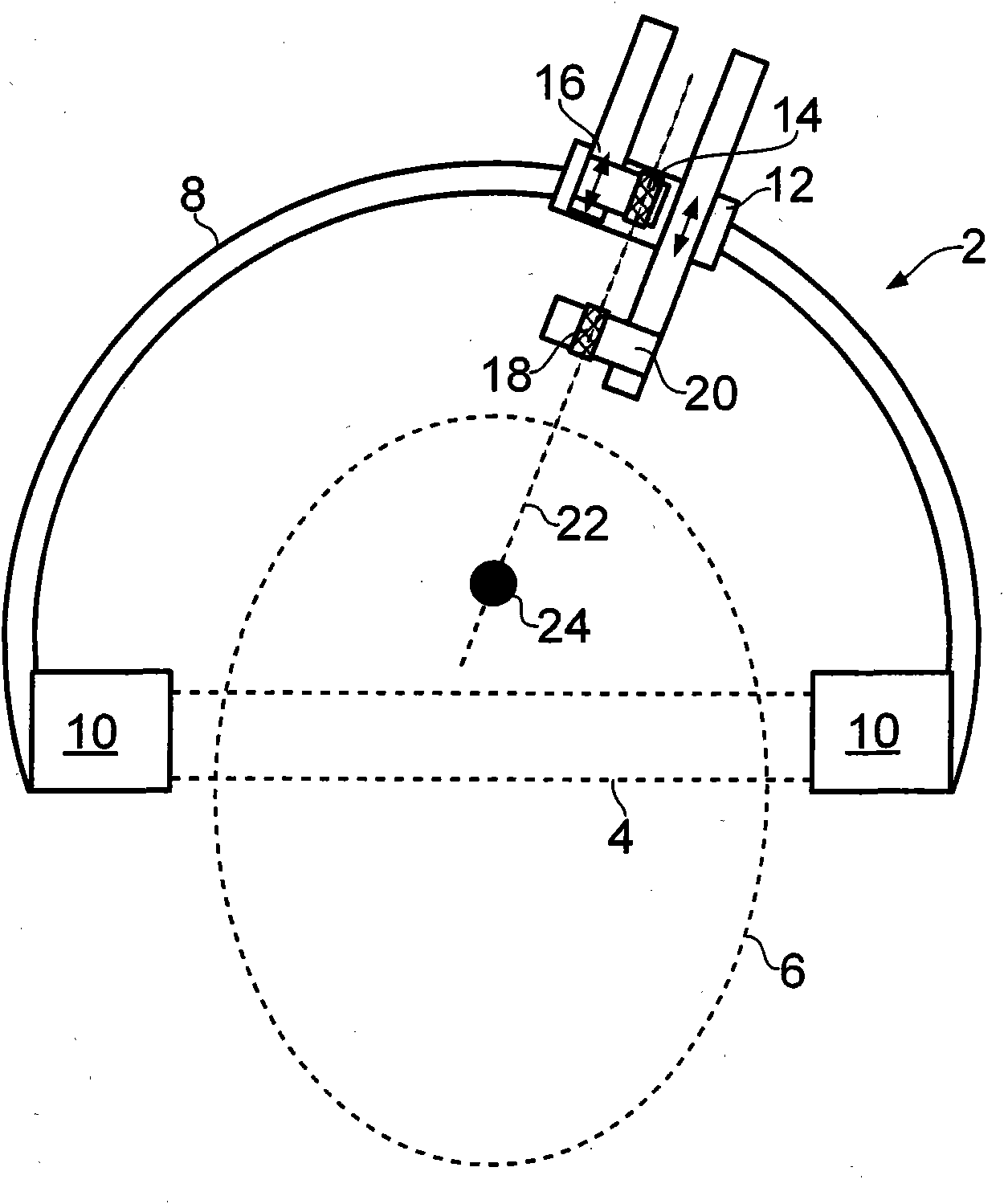
[0069] To perform neurosurgery, the surgeon first identifies the location of one or more desired targets in the brain. Stereotactic targeting of one or more targets in the brain may be performed by securely affixing a stereotaxic base ring to the subject's skull and using imaging techniques such as magnetic resonance imaging (MRI) to identify the location of the target or targets in the brain. position. The location of the target can be identified in three-dimensional coordinates by taking measurements against radiopaque fiducials attached to the stereotaxic base ring at known locations. The radiopaque fiducials may be contained in a device called a localization cassette that is reproducibly mounted on the stereotaxic base ring.

[0070] After obtaining the required MRI data, the positioning cassette can be removed from the stereotaxic base ring, which remains attached to the patient. The stereoguide can then be attached to the stereotaxic base ring and used as a platform fr...


PUM


Abstract

A skull mount (50;150;170;200;300) is described that is attachable to a hole (60) formed in the skull. The skull mount (50;150;170;200;300) comprises an alignment guide (62;152;172;216;306) defining an alignment axis (22;210;312) along which neurosurgical instruments can be passed. The skull mount, when attached to a hole in a skull, is arranged such that it does not substantially protrude from the outermost surface of the skull and does not extend into the brain parenchyma. Also described is a neurosurgical alignment instrument (30,206) for aligning such a skull mount (50;150;170;200;300) that comprises an elongate shaft (32) and an element (34,36) protruding from the distal end of the elongate shaft (32) for engaging and aligning the alignment guide (62;152;172;216;306) of an associatedskull mount (50;150;170;200;300). When the alignment instrument is engaged with a skull mount attached to a hole formed in the skull, the protruding element passes through the alignment guide of the skull mount and into the cortex of the subject's brain.

Description

technical field [0001] The present invention relates to instruments used in neurosurgery and to neurosurgery methods. In particular, the present invention relates to apparatus and methods for use in stereotaxic targeted therapy of abnormal brain function, and apparatus and methods for correctly guiding the apparatus directly into the brain parenchyma. Background technique [0002] There are many situations in which it is desirable to deliver a therapeutic agent via an implanted catheter to a specific target within the brain parenchyma. Moreover, many of these therapeutic agents can cause unwanted side effects if delivered to healthy parts of the brain. Examples of treatment of abnormal brain function include acute infusion of gamma-aminobutyric acid agonists into epileptic foci or pathways to prevent dissemination, and chronic delivery of opiates or other analgesics to periaqueductal gray matter or thalamic targets for treatment unmanageable pain. Furthermore, cytotoxic a...

Claims


Application Information

Patent Timeline
Patent Type & Authority Applications(China)
IPC IPC(8): A61B19/00A61B17/34
CPCA61B17/3403A61B2019/208A61B19/201A61B2017/3407A61B17/3423A61B17/3415A61B2090/103A61B90/11
Inventor 雨果·乔治·戴瑞克保罗·大卫·菲尔德史蒂文·斯特里特菲尔德·吉尔
Owner RENISHAW (IRELAND) LTD
Learn More