Ultrasonic diagnosis apparatus, ultrasonic image processing apparatus, ultrasonic image processing method, and ultrasonic image processing program

A diagnostic device and image processing technology, applied in ultrasonic/sonic/infrasonic diagnosis, sonic diagnosis, infrasonic diagnosis and other directions, can solve the problems of difficulty in observing the placenta, difficulty in observing blood vessels in the superficial part of the placenta, etc., and achieve the effect of improving the accuracy of image diagnosis.

Active Publication Date: 2011-04-27
TOSHIBA MEDICAL SYST CORP
View PDF8 Cites 16 Cited by
  • Summary
  • Abstract
  • Description
  • Claims
  • Application Information

AI Technical Summary

Problems solved by technology

Therefore, observation of blood vessels on the surface of the placenta becomes difficult
In addit

Method used


Image

  • Ultrasonic diagnosis apparatus, ultrasonic image processing apparatus, ultrasonic image processing method, and ultrasonic image processing program
  • Ultrasonic diagnosis apparatus, ultrasonic image processing apparatus, ultrasonic image processing method, and ultrasonic image processing program
  • Ultrasonic diagnosis apparatus, ultrasonic image processing apparatus, ultrasonic image processing method, and ultrasonic image processing program

Examples


Embodiment Construction

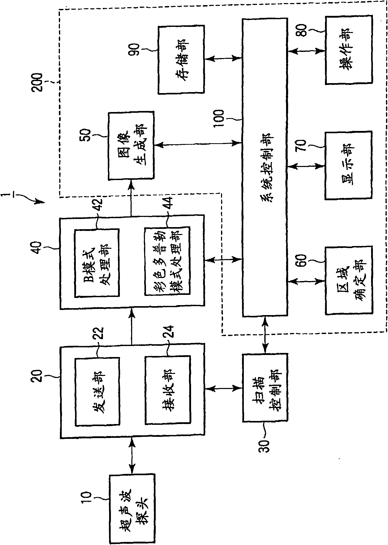
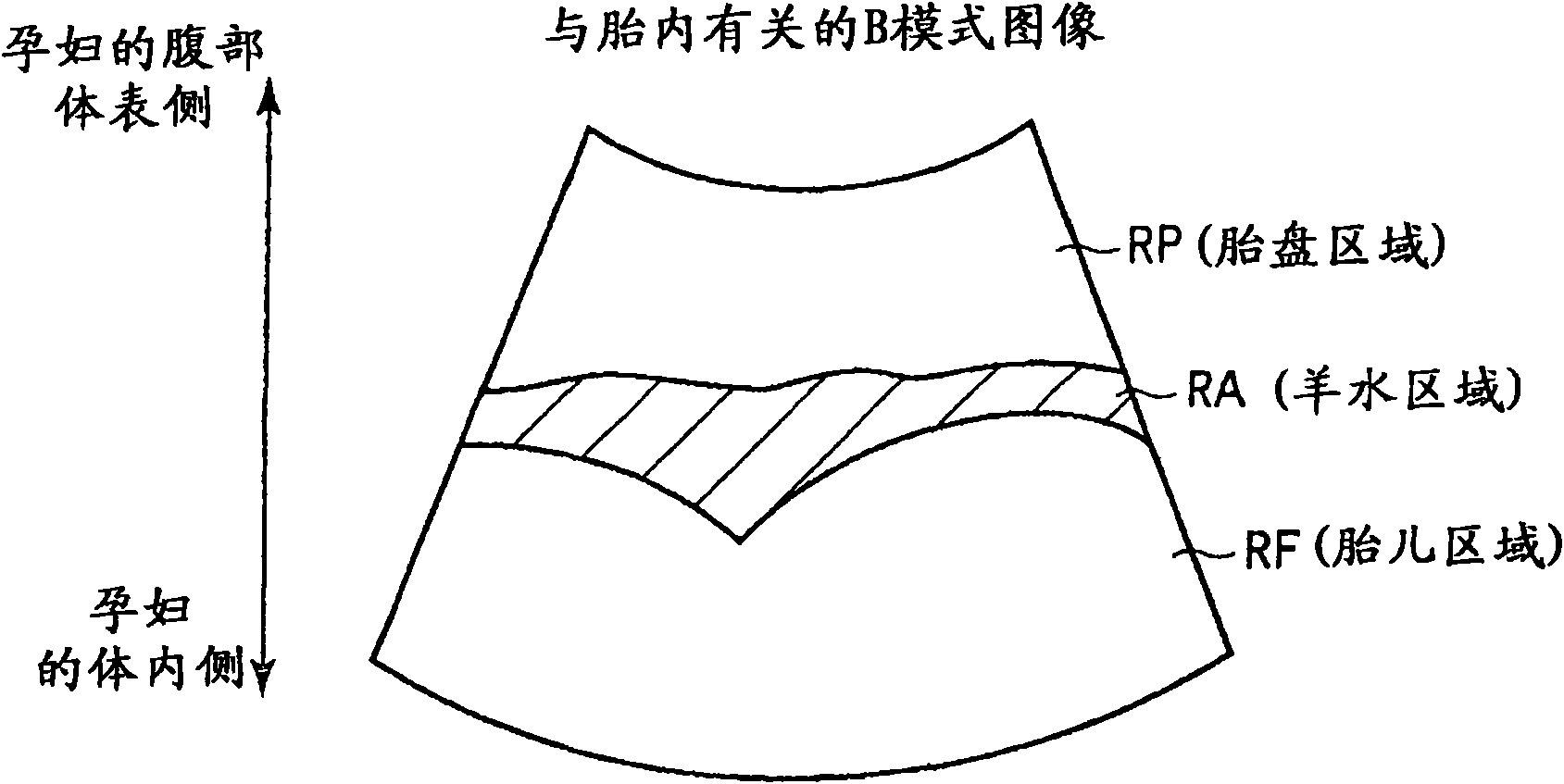
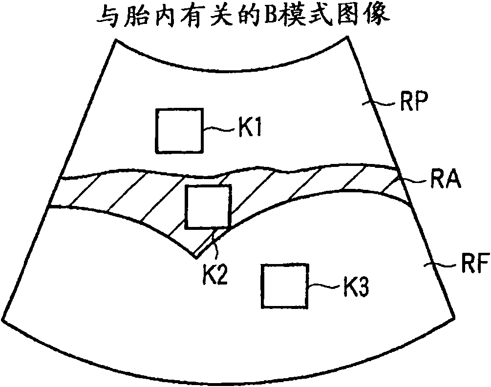
[0023] The ultrasonic diagnostic apparatus according to the present embodiment executes B-mode scanning and color Doppler mode scanning via an ultrasonic probe on a scanning area related to the inside of a pregnant woman's womb. The ultrasonic diagnostic apparatus according to the present embodiment includes a first generating unit, a specifying unit, a second generating unit, and a display unit. The first generation unit generates first color Doppler mode data related to the scanning region based on the output from the ultrasound probe during the color Doppler mode scan, The output of the probe generates the 1st B-mode data related to the above-mentioned scanning area. The specifying unit specifies a specific region including at least one of an amniotic fluid region and a fetal region based on the signal intensity distribution or brightness distribution of the first B-mode data. The second generating unit generates second color Doppler pattern data in which data correspondin...


PUM


Abstract

An ultrasonic diagnosis apparatus executes B-mode scanning and color Doppler mode scanning on a scanning region associated with an interior of the womb of a pregnant woman via an ultrasonic probe. A first generating unit generates first color Doppler mode data and generates first B-mode data. A specifying unit specifies a specific region including at least one of an amniotic fluid region and a fetus region based on one of a signal intensity distribution and a luminance distribution of the first B-mode data. A second generating unit generates second color Doppler mode data by deleting a specific data from the first color Doppler mode data. The specific color Doppler mode data corresponds to the specific region. A display unit displays a color Doppler mode image and a first B-mode image while superimposing the color Doppler mode image and the first B-mode image.

Description

[0001] (Cross-reference to related application) [0002] This application is based on and claims priority from prior Japanese Patent Application No. 2009-234271 filed on October 8, 2009, the entire contents of which are hereby incorporated by reference. technical field [0003] The present embodiment relates to an ultrasonic diagnostic apparatus, an ultrasonic image processing apparatus, an ultrasonic image processing method, and an ultrasonic image processing program. Background technique [0004] The color Doppler mode image is an image in which fluid information such as a velocity component, a power component, and a variance component of a flowing object such as a blood flow are expressed in color by the color Doppler method. During ultrasonography, a color Doppler mode image is displayed superimposed on a B-mode image. [0005] These techniques are used to perform ultrasound examinations of pregnant women's fetuses. As one of the applications of the ultrasonography met...

Claims


Application Information

Patent Timeline
IPC IPC(8): A61B8/06
CPCA61B8/467A61B8/13A61B8/469A61B8/0866A61B8/463A61B8/06
Inventor 浜田贤治岭喜隆
Owner TOSHIBA MEDICAL SYST CORP
Why Eureka
Try Eureka