System and method for imaging with enhanced depth of field

A technology of imaging and imaging devices, applied in the generation of 2D images, image data processing, details involving computational photography, etc., can solve problems such as unsatisfactory artifacts

Inactive Publication Date: 2011-05-11
GENERAL ELECTRIC CO
View PDF7 Cites 15 Cited by
  • Summary
  • Abstract
  • Description
  • Claims
  • Application Information

AI Technical Summary

Problems solved by technology

Unfortunately, the use of these techniques introduces unsatisfactory artifacts in the resulting image
Furthermore, these techniques tend to produce images with limited focus quality (especially when faced with samples mounted on glass slides that are not sufficiently flat within a single field of view), thereby limiting the use of these microscopes in pathology laboratories for diagnosis Abnormalities in such samples (especially where the diagnosis requires high magnification) (eg, with respect to bone marrow aspirate)

Method used


Image

  • System and method for imaging with enhanced depth of field
  • System and method for imaging with enhanced depth of field
  • System and method for imaging with enhanced depth of field

Examples


Embodiment Construction

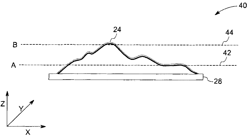
[0019] As will be described in detail below, methods and systems are provided for imaging samples such as samples with appreciable materials in the non-slide plane, while enhancing image quality and optimizing scanning speed. By adopting the methods and devices described below, enhanced image quality and a considerably increased scanning speed can be obtained, while simplifying the clinical workflow of sample scanning.

[0020] Although, the exemplary embodiments illustrated in the following are described in the context of a digital microscope, it will be appreciated that imaging devices are used in, for example, but not limited to, telescopes, cameras, or medical scanners (such as X-ray computed tomography (CT) imaging systems, etc. ) And other applications are also considered in conjunction with this technology.

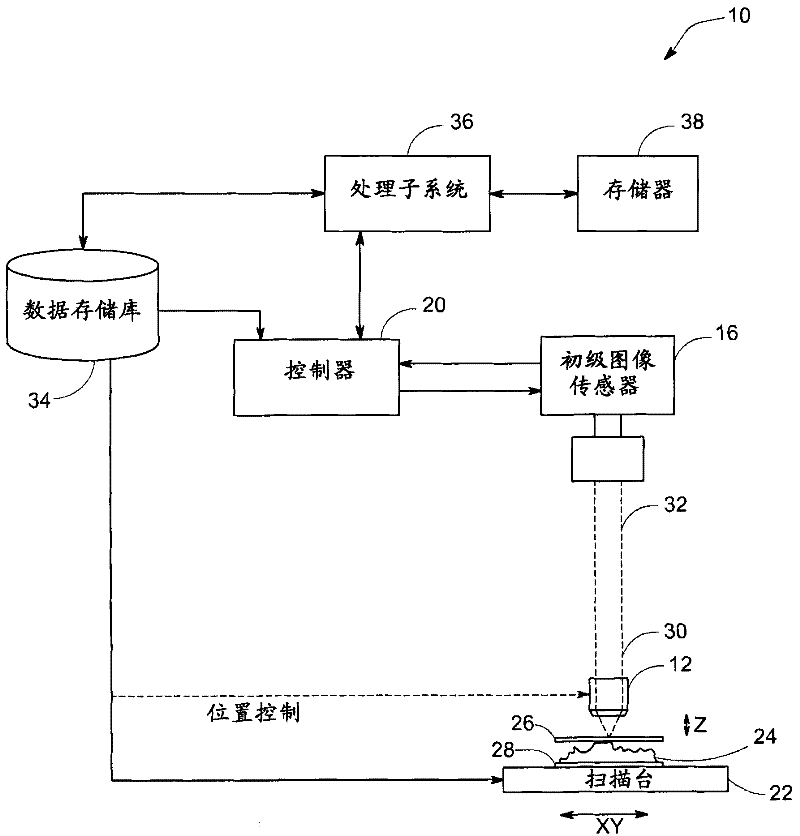
[0021] figure 1 Illustrated is an embodiment of an imaging device 10, such as a digital optical microscope, which includes aspects of the present invention. The imagin...


PUM


Abstract

A method for imaging is presented. The method includes acquiring a plurality of images corresponding to overlapping fields of view at a plurality of sample distances using an imaging device having an objective and a stage for holding a sample to be imaged. Moreover, the method includes determining a figure of merit corresponding to each pixel in each of the plurality of acquired images. The method also includes synthesizing a composite image based upon the determined figures of merit.

Description

Technical field [0001] Embodiments of the invention relate to imaging, and more specifically to the construction of images with enhanced depth of field. Background technique [0002] The prevention, monitoring, and treatment of physiological conditions such as cancer, infectious diseases and other diseases require timely diagnosis of these physiological conditions. Generally, biological samples from patients are used for disease analysis and identification. Microscopic analysis is a technique widely used in the analysis and evaluation of these samples. More specifically, samples can be studied to detect the presence of abnormal numbers or types of cells and / or tissues that can be indicative of a disease state. Automated microscope analysis systems have been developed to facilitate rapid analysis of these samples and have the advantage of accuracy over manual analysis in which technicians may become fatigued over time leading to misreading of the samples. Typically, a sample on...

Claims


Application Information

Patent Timeline
Patent Type & Authority Applications(China)
IPC IPC(8): G02B21/36G02B27/00G01N21/84
CPCG02B21/367G06T11/00G06T2200/21
Inventor K·B·肯尼D·L·亨德森
Owner GENERAL ELECTRIC CO
Why Eureka
Try Eureka