Endoscope system, method of using the same, assisting tool and adapter
A technology for endoscopes and adapters, applied in the field of endoscope systems, can solve problems such as multifunctional limitations
- Summary
- Abstract
- Description
- Claims
- Application Information
AI Technical Summary
Problems solved by technology
Method used
Image
Examples
no. 1 approach
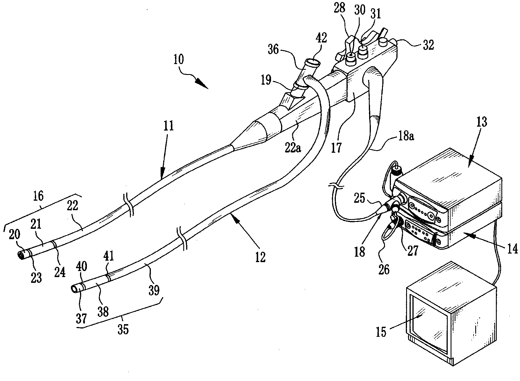
[0100] refer to figure 1 , The endoscope system 10 includes a nasal endoscope (hereinafter referred to as endoscope) 11 , an assisting tool 12 , a light source device 13 , a processing device 14 and a monitor 15 . The endoscope 11 has an insertion portion 16 to be inserted through one of the patient's external nostrils. The insertion portion 16 is connected to the operation portion 17 through the grip portion 22a. The operation section 17 is connected to a universal cable 18a. At the front end of the universal cable 18a, there is provided a universal connector 18 that connects the endoscope 11, the light source device 13 and the processing device 14 together.
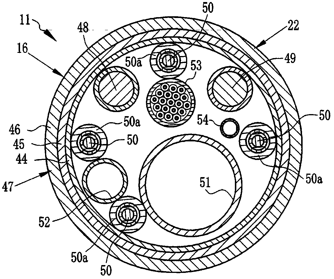
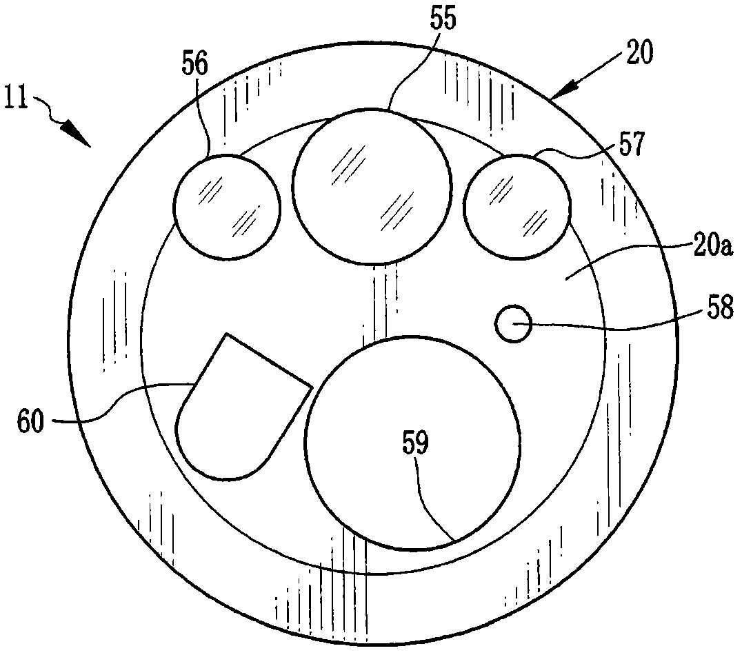
[0101] The insertion portion 16 is substantially hollow along its entire length and contains the tweezers channel. The tweezers channel is connected at one end to the tweezers outlet port on the front of the insertion portion 16 and at the other end to the tweezers inlet port 19 on the grip 22a. A tweezers entry por...
no. 2 approach
[0156] Next, another embodiment of the present invention is described. Hereinafter, elements similar to those of the first embodiment are denoted by the same reference numerals, and their detailed descriptions are omitted.
[0157] Such as Figure 16 As shown, the endoscope system 160 includes an endoscope 161 , an assisting tool 162 , a light source device 163 , a processing device 164 and a monitor 165 .
[0158] The auxiliary tool 162 has an insertion portion 35 and an auxiliary LG connector 166 . The auxiliary LG connector 166 is provided at the front end of the cord 168 extending from the proximal end portion 167 of the insertion portion 35 , and is detachably connected to the auxiliary LG socket 170 of the light source device 163 .
[0159] The insertion portion 35 of the assisting tool 162 comprises a light guide extending from the distal end 37 to the proximal end 167 in the inner space. Such a light guide transmits the illumination light emitted from the light sour...
no. 3 approach
[0202] Described below is another preferred embodiment of the present invention with magnets of different forms.
[0203] Such as Figure 31 As shown, an endoscope system 300 includes an endoscope 301 for capturing images inside a patient, an assisting tool 302 for providing additional functions to the endoscope 301, and an auxiliary tool 302 for providing illumination light to the endoscope 301 to illuminate the patient. A light source device 303 inside the body, a processing device 303 for generating an endoscopic image, and a monitor 305 for displaying the endoscopic image.
[0204] This endoscope 301 includes an insertion part 306 to be inserted into the patient's body, an operation part 307 connected to the proximal end of the insertion part 306 and operated by an operator such as a doctor or a technician, and an operation part 307 connected to the operation part 307 to insert the endoscope The mirror 301 is connected to the light source device 303 and the processing dev...
PUM
 Abstract
Description
Claims
Application Information
 - R&D
- Intellectual Property
- Life Sciences
- Materials
- Tech Scout
- Unparalleled Data Quality
- Higher Quality Content
- 60% Fewer Hallucinations
Browse by: Latest US Patents, China's latest patents, Technical Efficacy Thesaurus, Application Domain, Technology Topic, Popular Technical Reports.
© 2025 PatSnap. All rights reserved.Legal|Privacy policy|Modern Slavery Act Transparency Statement|Sitemap|About US| Contact US: help@patsnap.com


