
Penetrating endoscope and endoscopic surgical instrument with CMOS image sensor and display

a technology of endoscope and image sensor, which is applied in the field of endoscopes, can solve the problems of inconvenient use, inconvenient sterilization and maintenance, and inability to accurately measure the size of the endoscope, and achieve the effects of reducing the cost of sterilization and maintenance, and improving the accuracy of the endoscop

Inactive Publication Date: 2002-05-14
YOON INBAE
View PDF15 Cites 317 Cited by
  • Summary
  • Abstract
  • Description
  • Claims
  • Application Information

AI Technical Summary

Problems solved by technology

CCD solid-state image sensors provide the benefits of small size and flexibility in image sensor placement, but are relatively difficult and expensive to implement in medical instruments, since CCD sensor signals require processing through complex and expensive ancillary equipment, if a usable image is to be displayed.
Endoscopic instruments, in general, also have been so expensive to use, sterilize and maintain that only surgeons in well-funded medical facilities have had access to instruments providing visualization in the body.
Prior art endoscopes also suffer from many disadvantages when used in the procedures for which they are presently recommended.
A major disadvantage of prior art endoscopes is that they cannot be used during penetration of anatomical tissue to view the anatomical tissue being penetrated (e.g. using a trocar penetrating member).
Thus, it is difficult for a surgeon to know whether to alter the path of the perietrating member to avoid blood vessels and other types of anatomical tissue and organs.
While the Visiport m and Optiview.TM. optical trocars do provide visibility during the penetrating step, they are still subject to the disadvantages associated with conventional endoscopic surgical procedures as outlined above and so are not useful in moving patient care out of expensive OR facilities and closer to the bed side.

Method used


Image

  • Penetrating endoscope and endoscopic surgical instrument with CMOS image sensor and display
  • Penetrating endoscope and endoscopic surgical instrument with CMOS image sensor and display
  • Penetrating endoscope and endoscopic surgical instrument with CMOS image sensor and display

Examples


third embodiment

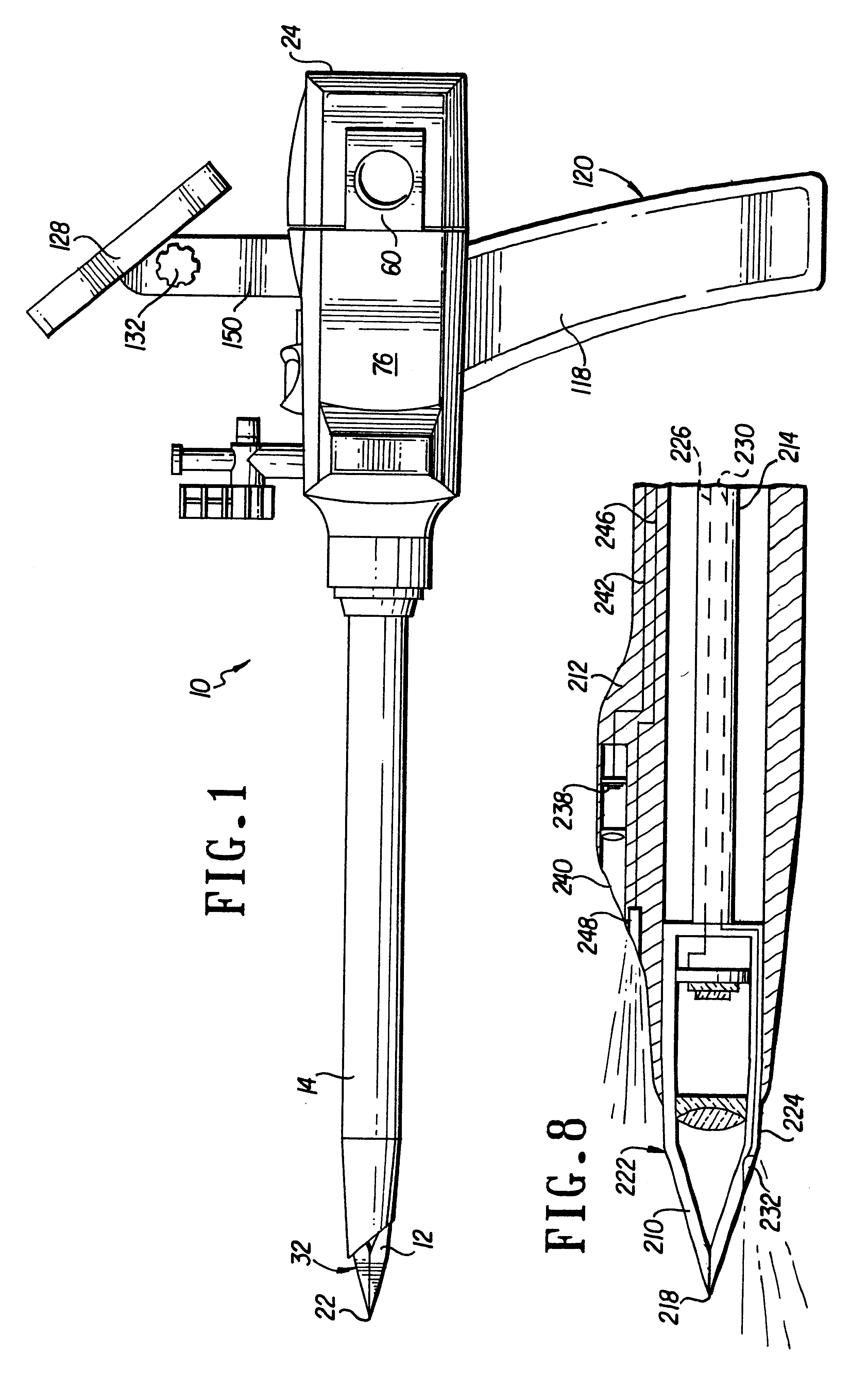
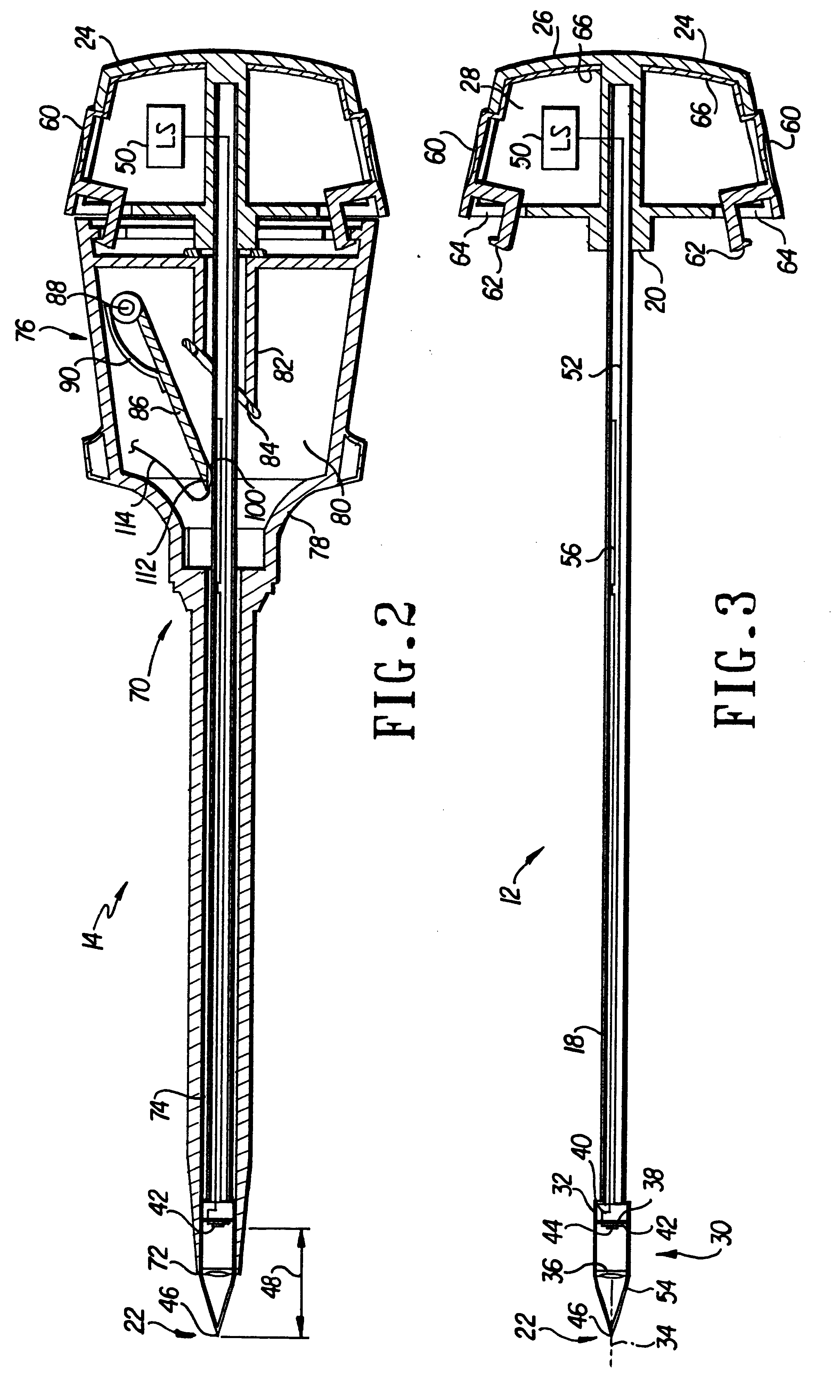
Turning now to FIGS. 15, 16 and 17, the penetrating endoscope 200 (FIGS. 7 and 8) is illustrated. As shown in FIG. 15, penetrating endoscope 200 is positioned against the outer tissue wall 260 in preparation for penetrating through the tissue wall using sharp distal end 218 containing penetrating member CMOS image sensor 220, the view from which is shown on display 243. The surgeon grasps handle 237 and applies sufficient force to the penetrating endoscope 200 to force the sharp distal end through outer tissue wall 260 while viewing the field of view of the penetrating member image sensor 222 on display 243. Once the surgeon has forced sharp distal end 218 through outer tissue wall 260, the interior of the body and, more particularly, interior organ wall 262 are illuminated and become visible to both the penetrating member image sensor 22 and the portal sleeve image sensor 238.

In the method of the present invention, display 243 receives image signal information from the penetrating ...

first embodiment

The endoscopic instrument according to the present invention is illustrated in FIGS. 18 and 19 and includes an elongate cylindrical barrel, or outer shaft 312 with a longitudinal axis and preferably having one or more elongated lumens or passages defined therein (preferably in the form of one or more channels, e.g., 314), a barrel distal end 315 for being disposed in the body and a barrel proximal end terminating in and carried by a housing 318 including an exterior wall enclosing an interior volume. Housing 318 includes scissor type handles 320 and 322 for controlling surgical instruments such as forceps having jaw members 324, 325 (as best seen in FIG. 19), or other end effectors. Housing 318 also includes a transversely located button 326 for selectively disengaging the scissor type handles 320 and 322 and permitting rotation of the handles about the axis of rotation (as indicated by the arrow A in FIG. 18). Handles 320, 322 are connected through a linkage mechanism (as is well k...


PUM


Abstract

A penetrating endoscope provides visualization of organ or tissue structures or foreign objects in a body. The penetrating endoscope includes an elongate penetrating member, a complementary metal dioxide semiconductor (CMOS) image sensor and an objective lens. The CMOS image sensor is substantially planar and includes a plurality of pixels with a pixel signal processing circuit for generating a color image ready signal. The CMOS image sensor converts image light energy into electrical color image ready signal energy for transmission out of the body. The color image ready signal is viewed on a color image display. The CMOS image sensor is carried on the elongate penetrating member adjacent a distal end of the elongate penetrating member. The objective lens is also carried on the distal end of the elongate penetrating member on an optical axis and focuses an image corresponding to an endoscope field of view at an image plane intersecting the optical axis. The CMOS image sensor is mounted with the CMOS image sensor pixels disposed substantially in the image plane and on the optical axis. The penetrating endoscope may include end effectors such as cutters and forceps.

Description

This application claims benefit of U.S. application Ser. No. 60 / 085,243, filed May 13, 1998, which claim benefit of U.S. application Ser. No. 60 / 085,242, which claim benefit of U.S. application Ser. No. 60 / 093 / 069, filed Jul. 16, 1998.1. Field of the InventionThe present invention pertains to endoscopes in general and, more particularly, to a penetrating endoscopic instrument with a solid-state image sensor for penetrating into anatomical tissue and an endoscopic surgical instrument carrying an endoscope with a solid-state image sensor.2. Discussion of the Prior ArtVarious procedures are accomplished in both open surgery and endoscopic surgery, and generally include multiple steps requiring various operating instruments. "Open surgery" refers to surgery wherein the surgeon gains access to the surgical site by a relatively large incision and "endoscopic surgery" refers to minimally invasive surgery wherein the surgeon gains access to the surgical site via one or more portals through ...

Claims


Application Information

Patent Timeline
IPC IPC(8): A61B1/05A61B1/313A61B1/00A61B17/34A61B1/04A61B19/00H04N5/225
CPCA61B1/00052A61B1/00087A61B1/00135A61B1/00181A61B1/3132A61B17/3494A61B1/05H04N2005/2255A61B19/5225A61B90/37H04N23/555
Inventor YOON, INBAE
Owner YOON INBAE
Learn More