Real-time medical ultrasonic three-dimensional imaging method

A three-dimensional imaging and ultrasound technology, applied in medical science, ultrasound/sonic/infrasonic diagnosis, sound wave diagnosis, etc., can solve problems such as intersecting positions, achieve the effects of improving efficiency, solving memory bandwidth, and reducing useless redundant calculations

Inactive Publication Date: 2011-11-23
SHENZHEN LANDWIND IND
View PDF8 Cites 14 Cited by
  • Summary
  • Abstract
  • Description
  • Claims
  • Application Information

AI Technical Summary

Problems solved by technology

[0009] The present invention focuses on solving the problem of three-dimensional data reconstruction and visualization using the ray projection method
[0010] The invention uses the method of morphology to replace the traditional geometric analysis operation, and quickly solves the problem of the intersection position of the projection light and the volume data

Method used


Image

  • Real-time medical ultrasonic three-dimensional imaging method
  • Real-time medical ultrasonic three-dimensional imaging method
  • Real-time medical ultrasonic three-dimensional imaging method

Examples


Embodiment Construction

[0066] Below according to accompanying drawing and embodiment the present invention will be described in further detail:

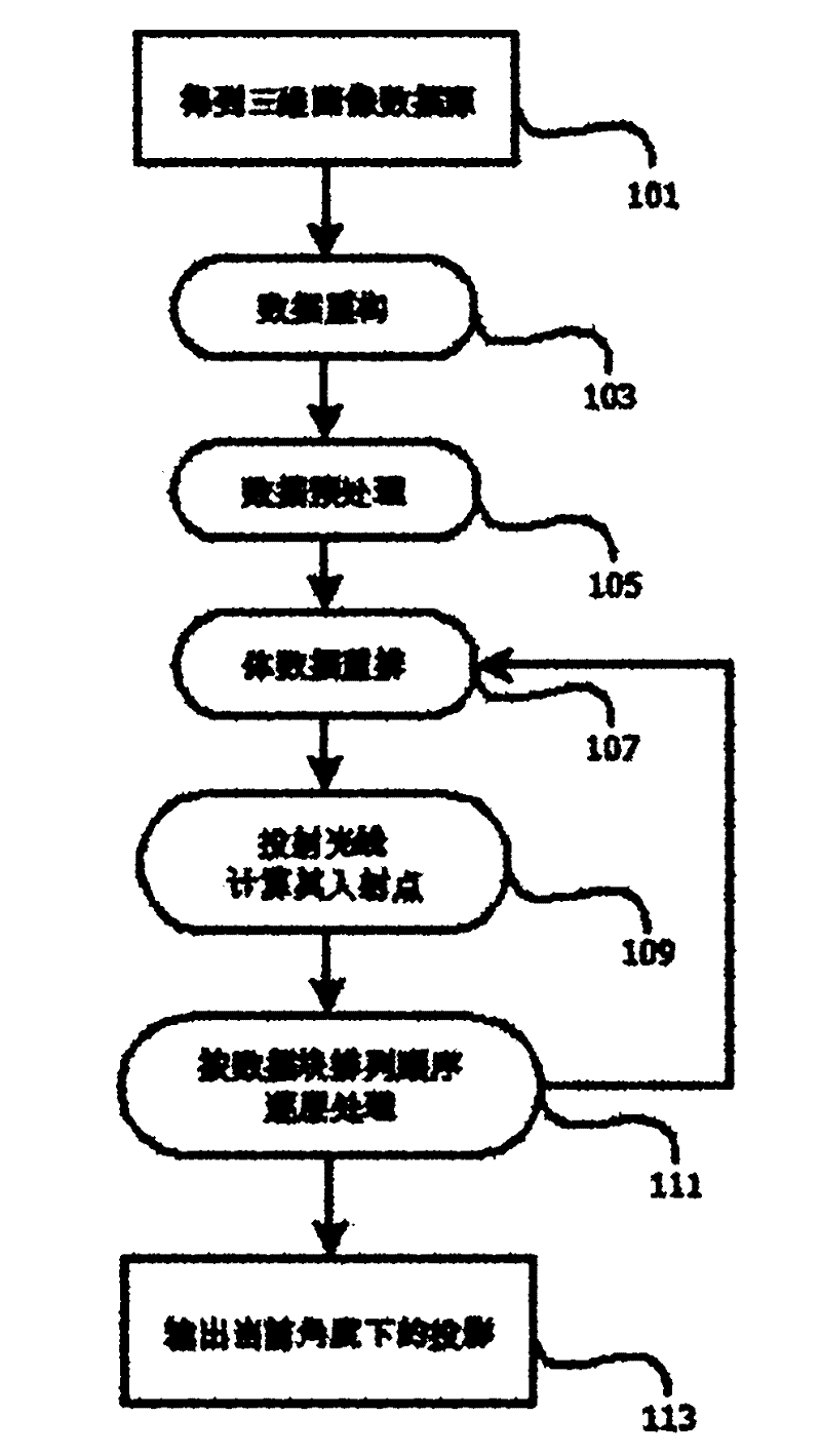
[0067] figure 1 It is a flow chart of real-time medical three-dimensional ultrasonic imaging implemented by the invention, which simply describes the medical three-dimensional ultrasonic real-time imaging processing flow. This processing method is applicable to medical three-dimensional ultrasonic images, and has application value in medical three-dimensional imaging systems such as CT and MRI.

[0068] The present invention considers and focuses on solving the problem of fast realization of three-dimensional data reconstruction and visualization. Therefore, the present invention firstly solves the problem of three-dimensional data storage. In order to express enough information, the capacity of three-dimensional data is usually quite large. At present, the storage method of CPU memory is one-dimensional. If we simply follow the general image storage met...


PUM


Abstract

The invention provides a real-time medical ultrasonic three-dimensional imaging method, which is applicable to real-time acquisition of three-dimensional medical ultrasonic volume data which may be stored. Aiming at the characteristics of a current central processing unit (CPU) and the characteristics of a three-dimensional ultrasonic image, storage and access modes of three-dimensional data are optimized, and the conventional ray casting method is improved in combination with the knowledge of image morphology, so that the reconstruction and visualization of the three-dimensional data are quickly realized.

Description

technical field [0001] The invention relates to rapid reconstruction and visualization of three-dimensional medical images. Background technique [0002] The waveform diagrams and two-dimensional cross-sectional diagrams that traditional medical diagnosis and treatment rely on are not intuitive enough for observation. In this regard, Derek Ney formally proposed the concept of medical visualization for the first time in the SIGGRAPH Course in 1994, that is, transforming two-dimensional images into more intuitive and realistic three-dimensional images, which can be observed, analyzed and interacted from multiple angles and levels Operation, providing a safer, more reliable and accurate solution for medical diagnosis and treatment. [0003] Usually, according to the method of data description in the rendering process, the visualization of medical volume data can be divided into three categories: one is to describe the three-dimensional structure of the object by splicing and f...

Claims


Application Information

Patent Timeline
Patent Type & Authority Applications(China)
IPC IPC(8): G06T15/06G06T17/00A61B8/00
Inventor 潘梁亮刘龙陆汇海徐漫涛张羽
Owner SHENZHEN LANDWIND IND
Why Eureka
Try Eureka