Intracavitary stent for medical radiotherapy and chemotherapy
An inner stent and cavity technology, applied in the field of medical radiotherapy and chemotherapy cavity stent, can solve the problems of ineffective treatment, life-threatening, easy perforation, etc., to improve the local control rate, prolong the survival period, and affect the normal tissue. less effect
- Summary
- Abstract
- Description
- Claims
- Application Information
AI Technical Summary
Problems solved by technology
Method used
Image
Examples
Embodiment 1
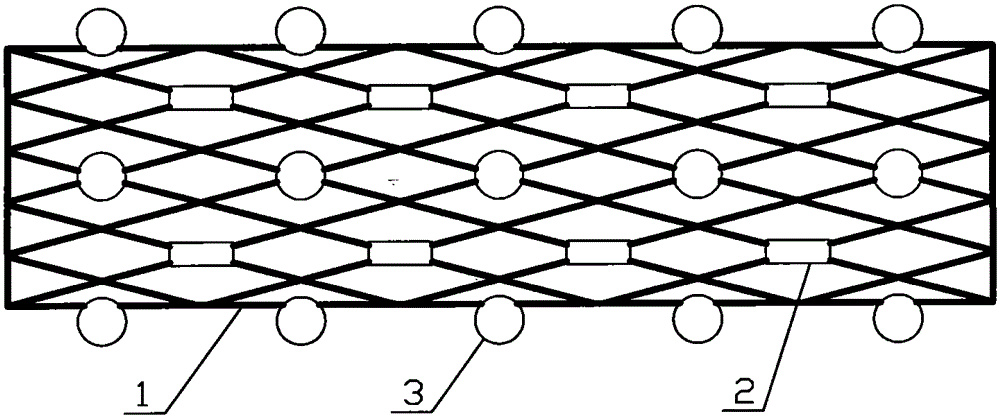
[0027] Embodiment 1: as figure 1 As shown, a stent in a medical radiotherapy and chemotherapy cavity includes a stent main body 1, 16 airtight radiotherapy chambers 2 are fixedly arranged on the outer wall of the stent main body 1, and radiotherapy particles are arranged in the radiotherapy chamber 2; The gap is 1.5-2.5 cm; chemotherapy drugs 3 are fixed on the outer wall of the bracket main body 1 between adjacent radiotherapy chambers 2 . Chemotherapeutic drugs 3 are bound on the stent main body 1 . Chemotherapy drug 3 is mitomycin C microspheres, or cisplatin microspheres, or doxorubicin microspheres, or 5-fluorouracil microspheres, or camptothecin microspheres, or methotrexate microspheres, or Any one of daunorubicin microspheres, or Brucea javanica oil microcapsules, or epinephrine gel, or carboplatin, or carmustine (BCNU), or paclitaxel, or doxorubicin.
[0028] The advantage of this embodiment is that the chemotherapeutic drug 3 and the radiotherapy chamber 2 are plac...
Embodiment 2
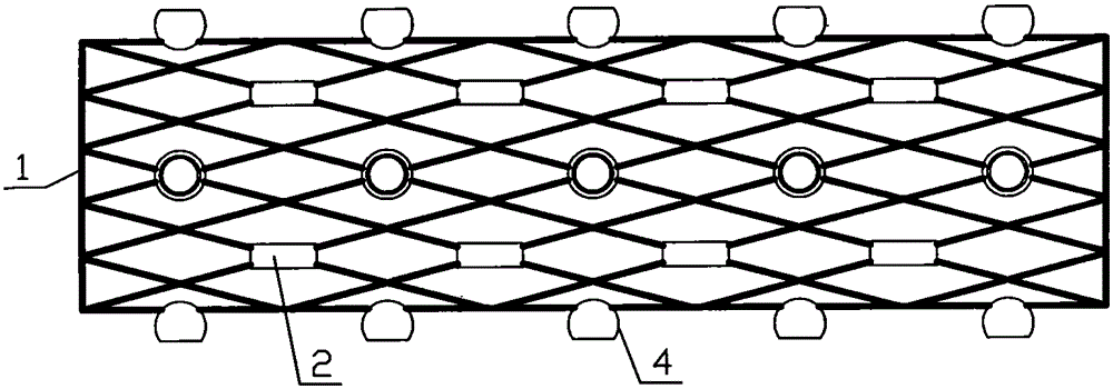
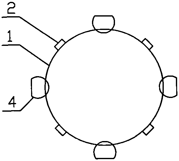
[0038] Embodiment 2: as figure 2 , image 3 and Figure 4 As shown, a stent in a medical radiotherapy and chemotherapy cavity includes a stent main body 1, 16 airtight radiotherapy chambers 2 are fixedly arranged on the outer wall of the stent main body 1, and radiotherapy particles are arranged in the radiotherapy chamber 2; The gap is 1.5 to 2.5 cm; chemotherapy drugs 3 are fixed on the outer wall of the stent body 1 between adjacent radiotherapy chambers 2, and the chemotherapeutic drugs 3 are placed in a spherical drug chamber 4, which is fixed in the adjacent radiotherapy chamber On the outer wall of the stent main body 1 between the 2, a drug release port 5 is provided on the outer wall of the spherical medicine compartment 4, and the drug release port 5 faces the outside of the stent main body 1. Chemotherapy drug 3 is mitomycin C microspheres, or cisplatin microspheres, or doxorubicin microspheres, or 5-fluorouracil microspheres, or camptothecin microspheres, or met...
Embodiment 3
[0049] Embodiment 3: as Figure 5 , Figure 6 and Figure 7 As shown, a stent in a medical radiotherapy and chemotherapy cavity includes a stent main body 1, 16 airtight radiotherapy chambers 2 are fixedly arranged on the outer wall of the stent main body 1, and radiotherapy particles are arranged in the radiotherapy chamber 2; The gap is 1.5-2.5 cm; chemotherapy drugs 3 are fixed on the outer wall of the stent body 1 between adjacent radiotherapy chambers 2,
[0050] Chemotherapeutic drugs 3 are placed in a medicine bag 6 with an opening at one end, and a tie rope 7 is provided on the medicine bag 6 , and the medicine bag 6 is bound to the outer wall of the bracket body 1 between adjacent radiotherapy chambers 2 through the tie rope 7 .
[0051] The medicine bag 6 is made of bioabsorbable fiber, such as polylactic acid (PDLLA, PLLA, PLA) material, or polyglutamic acid (polyglutamic acid PGA), or polylactic acid-glycolic acid copolymer (PLGA), or polydioxane Ketone (PPDO), ...
PUM
| Property | Measurement | Unit |
|---|---|---|
| porosity | aaaaa | aaaaa |
Abstract
Description
Claims
Application Information
 - R&D
- Intellectual Property
- Life Sciences
- Materials
- Tech Scout
- Unparalleled Data Quality
- Higher Quality Content
- 60% Fewer Hallucinations
Browse by: Latest US Patents, China's latest patents, Technical Efficacy Thesaurus, Application Domain, Technology Topic, Popular Technical Reports.
© 2025 PatSnap. All rights reserved.Legal|Privacy policy|Modern Slavery Act Transparency Statement|Sitemap|About US| Contact US: help@patsnap.com


