
Method and device for reducing scanning dose of X-ray

A scanning dose and X-ray technology, applied in computerized tomography scanners, echo tomography, etc., to achieve high accuracy and reliability, and reduce the scanning dose

Active Publication Date: 2015-06-03
SIEMENS SHANGHAI MEDICAL EQUIP LTD
View PDF4 Cites 0 Cited by
  • Summary
  • Abstract
  • Description
  • Claims
  • Application Information

AI Technical Summary

Problems solved by technology

However, the hundreds of images offline show that the object to be inspected is moving. Since the projection data and reconstructed images with motion artifacts are useless, the object to be inspected needs to be scanned again
In this way, the subject receives an unnecessary scan dose from a previous "useless" scan

Method used


Image

  • Method and device for reducing scanning dose of X-ray
  • Method and device for reducing scanning dose of X-ray
  • Method and device for reducing scanning dose of X-ray

Examples


Embodiment Construction

[0038] In order to make the purpose, technical solution and advantages of the present invention clearer, the following examples are given to further describe the present invention in detail.

[0039] The method and device for reducing X-ray scanning dose of the present invention will be described in detail below by taking CT equipment as an example.

[0040] For the current double-row CT, usually the time for the data acquisition system to rotate around the object to be checked synchronously is less than 1s, such as 0.6s, and the frequency of human body movement is usually once every 1s, so during the scanning process, not every time The scanned original projection data sets all contain motion data of the object to be checked. Unless the subject suffers from Parkinson's syndrome, the patient with this symptom has a very fast movement frequency and requires a high-end CT with a short scan time to scan it. If the original projection data sets contain motion data of the object t...


PUM


Abstract

The invention relates to the field of medical equipment, and particularly relates to a method for reducing the scanning dose of X-ray. The method comprises the steps of: scanning a to-be-detected object; detecting whether the to-be-detected object moves or not while scanning; and when the movement of the to-be-detected object is detected, closing an X-ray source. The invention further provides a device for reducing the scanning dose of X-ray. According to the method and the device, whether the to-be-detected object moves or not can be detected in real time, and the X-ray source can be closed or the scanning can be stopped immediately after detecting the movement, so that the scanning dose of X-ray accepted by the to-be-detected object is reduced.

Description

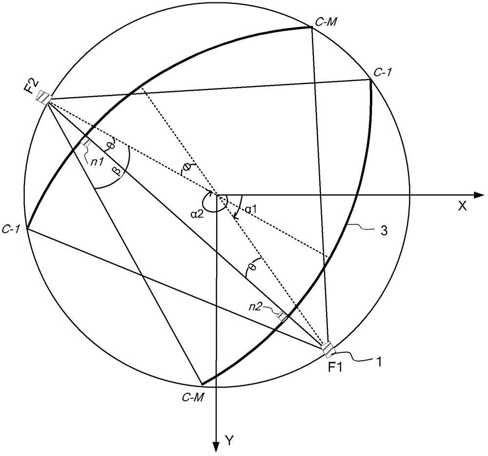
technical field [0001] The invention relates to the field of medical equipment, in particular to a method and device for reducing X-ray scanning dose. Background technique [0002] With the progress of medical level, X-ray computed tomography (Computed Tomography, CT) equipment has been widely used. When performing CT scanning on the object to be inspected, the rotating gantry rotates 360 degrees around the Z axis, and at the same time drives the data acquisition system including the X-ray source, collimator and detector to rotate 360° around the object to be inspected synchronously, so as to start from [0 , 360°] to acquire X-ray projection data, where the Z-axis is the horizontal direction of the CT examination bed entering and exiting the rotating gantry. At each projection angle, M channels of the detector simultaneously receive X-ray projection data and transmit it to the CT image reconstruction system for generating a reconstructed image. Since the human body inevita...

Claims


Application Information

Patent Timeline
Patent Type & Authority Patents(China)
IPC IPC(8): A61B6/03
Inventor 田毅杜春华王薇
Owner SIEMENS SHANGHAI MEDICAL EQUIP LTD
Learn More