Digital X-ray photography equipment, computed tomography equipment and related methods

A photographic equipment and tomography technology, applied in the field of medical imaging to avoid radiation doses

Pending Publication Date: 2020-03-10
SIEMENS SHANGHAI MEDICAL EQUIP LTD
View PDF8 Cites 0 Cited by
  • Summary
  • Abstract
  • Description
  • Claims
  • Application Information

AI Technical Summary

Problems solved by technology

The operator may enter and exit the scanning chamber several times until a satisfactory opening range is obtained

Method used


Image

  • Digital X-ray photography equipment, computed tomography equipment and related methods
  • Digital X-ray photography equipment, computed tomography equipment and related methods
  • Digital X-ray photography equipment, computed tomography equipment and related methods

Examples


Embodiment Construction

[0029] In order to make the purpose, technical solution and advantages of the present invention clearer, the following examples are given to further describe the present invention in detail.

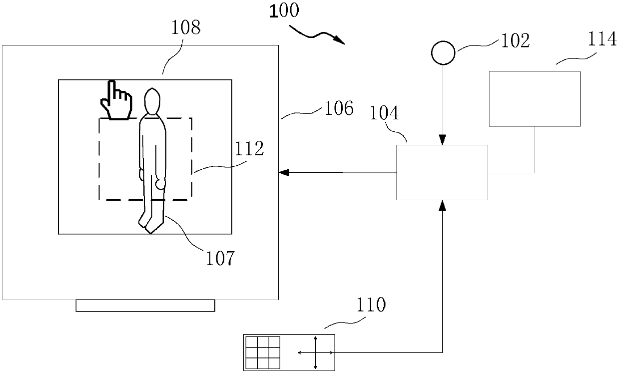
[0030] figure 1 It is a schematic diagram of a digital X-ray imaging device 100 according to the first embodiment of the present invention. Such as figure 1 As shown, the digital X-ray imaging device 100 includes an imaging unit 102 , a display unit 106 , an input unit 110 , a control unit 104 and a collimator 114 . The digital X-ray imaging device 100 may further include an illumination unit (not shown). The illumination unit projects light to the detector, which is used to indicate the projection range of X-rays on the detector (not shown) when the X-ray tube (not shown) is exposed. The inspected object 107 is located between the X-ray tube and the detector, and the imaging unit 102 takes an image of the area where the inspected object 107 is located. The display unit 106 displays ...


PUM


Abstract

The invention discloses digital X-ray photography equipment, a method for adjusting an opening range of a collimator of the digital X-ray photography equipment, computed tomography equipment and a method for determining a moving range of an examination table of the computed tomography equipment. The digital X-ray photography equipment includes a camera unit, a display unit, an input unit, a control unit and the collimator; the camera unit shoots images in a region where an examined object is located; the display unit displays video of the region provided by the camera unit; the input unit inputs a target range, corresponding to a part of the region, of the display unit; the control unit receives the target range from the input unit; and the collimator receives the target range from the control unit and adjusts the opening range accordingly. The digital X-ray photography equipment and related methods provided by the invention can conveniently adjust the opening range of the collimator and prevent an operator from entering and leaving a scanning room.

Description

technical field [0001] The invention relates to medical images, especially digital X-ray photography equipment and computer tomography equipment. Background technique [0002] Digital radiography equipment and computed tomography equipment are commonly used medical imaging equipment. [0003] Digital X-ray photography equipment has a collimator, which is used to limit the emission range of X-rays. Digital X-ray photography equipment is usually equipped with an illumination unit that projects light to the detector to indicate the projection range of X-rays on the detector when the X-ray tube is exposed. When in use, the operator needs to enter the scanning room and adjust the opening range of the collimator. The operator may enter and exit the scanning chamber several times until a satisfactory opening range is obtained. [0004] Computed tomography equipment usually needs to take positioning images before performing tomography. The operator determines the tomographic ran...

Claims


Application Information

Patent Timeline
IPC IPC(8): A61B6/03A61B6/06A61B6/04
CPCA61B6/03A61B6/06A61B6/0487
Inventor 赫伟刘颖慧
Owner SIEMENS SHANGHAI MEDICAL EQUIP LTD
Why Eureka
Try Eureka