
Method and device for dividing brain magnetic resonance angiography data

An angiography and magnetic resonance technology, applied in the field of image processing, can solve problems such as inability to segment blood vessels, and achieve the effect of reducing computing time

Active Publication Date: 2013-04-17
SHENZHEN INST OF ADVANCED TECH CHINESE ACAD OF SCI
View PDF2 Cites 6 Cited by
  • Summary
  • Abstract
  • Description
  • Claims
  • Application Information

AI Technical Summary

Problems solved by technology

Therefore, commonly used blood vessel segmentation methods cannot quickly and accurately segment blood vessels.

Method used


Image

  • Method and device for dividing brain magnetic resonance angiography data
  • Method and device for dividing brain magnetic resonance angiography data

Examples


Embodiment Construction

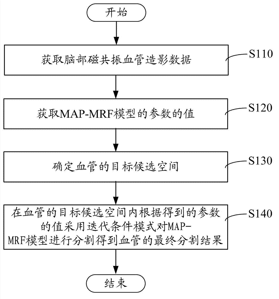
[0019] Please refer to figure 1 , one embodiment provides a method for segmenting brain magnetic resonance angiography data, the method for segmenting brain magnetic resonance angiography data includes the following steps:

[0020] Step S110, acquiring cerebral magnetic resonance angiography data.

[0021] Brain magnetic resonance angiography data were processed by multi-scale filtering method. To enhance blood vessels and suppress noise, get a normalized response R f , and R f ∈ [0, 1].

[0022] Step S120, acquiring the parameter values ​​of the MAP-MRF (maximum posteriori-Markov random field, maximum posteriori probability-Markov random field) model. The parameters here include mean, variance, and mixing ratio.

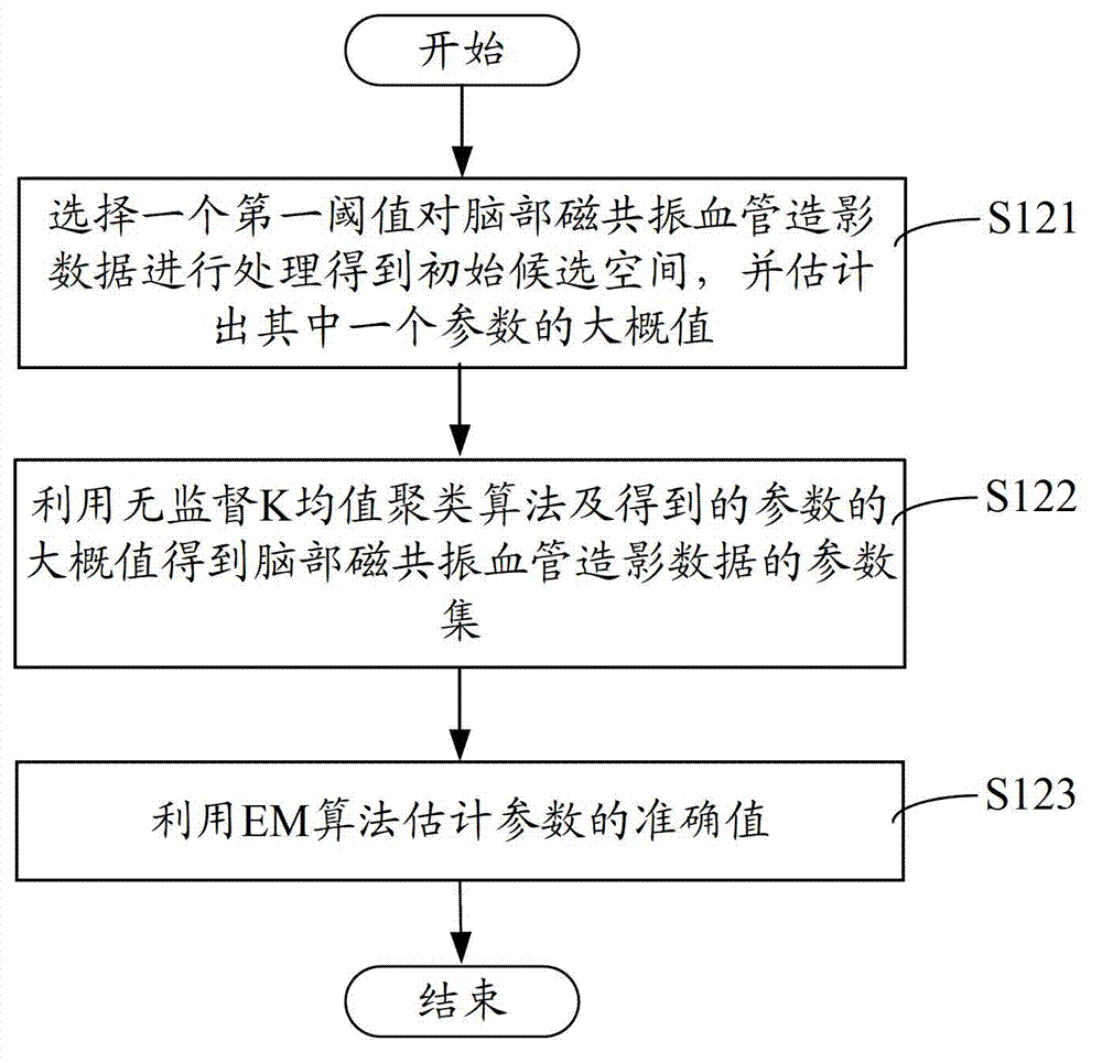
[0023] In step S120, obtaining the value of the parameter of the MAP-MRF model mainly includes the following steps.

[0024] Step S121, choose a first threshold γ 1 The initial candidate space of blood vessels is obtained by processing the brain magnetic reso...


PUM


Abstract

The invention provides a method and a device for dividing brain magnetic resonance angiography data. The method for dividing the brain magnetic resonance angiography data comprises the following steps of obtaining the brain magnetic resonance angiography data; obtaining values of parameters of an MAP-MRF (Maximum A Posteriori-Markov Random Field) model; determining target candidate space of a blood vessel; and dividing the MAF-MRF model in the target candidate space of the blood vessel according to the obtained values of the parameters by adopting an iterated conditional mode, and obtaining a final dividing result of the blood vessel. According to the method for dividing the brain magnetic resonance angiography data, disclosed by the invention, the dividing of the blood vessel is completed in the target candidate space, thus the calculating time is greatly reduced, and the brain blood vessel can be quickly divided.

Description

technical field [0001] The invention relates to an image processing method, in particular to a method and device for segmenting brain magnetic resonance angiography data. Background technique [0002] Every year, a large number of patients are affected by cerebrovascular diseases such as strokes caused by thrombosis, hemorrhage, arterial malformation, and stenosis. Therefore, accurate segmentation of cerebral vessels has important clinical significance for diagnosis, surgery, and preoperative planning. Due to the complex three-dimensional shape and fine structure of cerebral blood vessels, its image segmentation is very challenging. [0003] Magnetic resonance angiography (MRA, magnetic resonance angiography) is a non-intrusive flow imaging and three-dimensional volume data generation technique. There are many methods that can be used for MRA segmentation, and these methods can be divided into two categories: skeleton-based and non-skeleton methods. The former segment and...

Claims


Application Information

Patent Timeline
IPC IPC(8): G06T7/00
Inventor 周寿军胡庆茂谢耀钦辜嘉
Owner SHENZHEN INST OF ADVANCED TECH CHINESE ACAD OF SCI
Learn More