

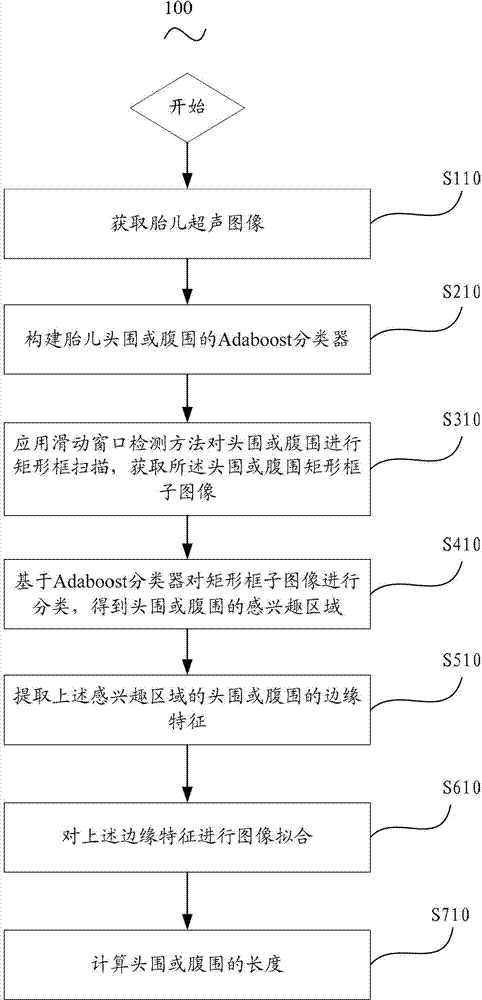
Measurement method for fetus ultrasound image
An ultrasound image and measurement method technology, which is applied in the field of fetal ultrasound image measurement, can solve the problems of low accuracy, lack of clinical application, and long measurement time, and achieves the effects of improving detection speed and accuracy and improving robustness.
- Summary
- Abstract
- Description
- Claims
- Application Information
AI Technical Summary
Problems solved by technology
Method used
Image

Examples
Embodiment
[0101] Experimental conditions: the experiment provided in this embodiment has a total of 675 sets of ultrasound head circumference images of fetuses. The data were provided by XXX Maternal and Child Health Hospital, and the ultrasonic diagnostic system was collected from Siemens Sequoia512 ultrasonic instrument. The fetal gestational age was distributed between 17 weeks and 38 weeks. Among them, 500 sets of fetal ultrasound images were used for training the Adaboost classifier of fetal head circumference, and the remaining 175 sets were used for measurement of fetal ultrasound images.
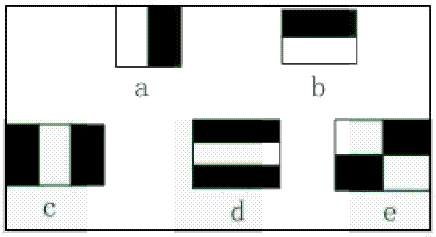
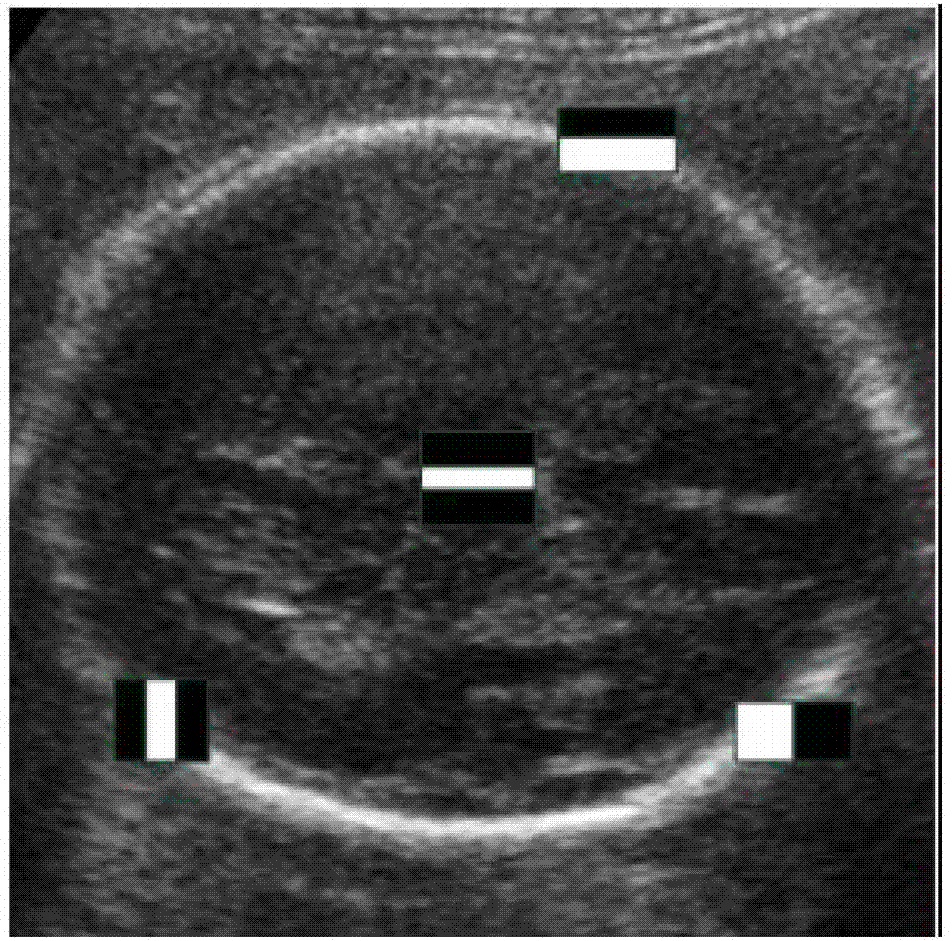
[0102] Automatic detection of regions of interest around the head circumference: The training samples consist of 500 training positive samples and 1200 randomly extracted negative samples near the positive samples. All samples are normalized to pixel size. We performed detection on 175 ultrasound images of head circumference. Comparing the experimental results with the clinician's standard ...
PUM
 Abstract
Description
Claims
Application Information