Endoscopic Nasopharyngeal Carcinoma Ultrasound Imaging Device

An ultrasonic imaging and nasopharyngeal cancer technology, applied in the field of medical devices, can solve the problems of inaccurate detection and achieve the effects of improved accuracy, low equipment maintenance costs, and low inspection costs

Active Publication Date: 2015-07-29
NAT INST OF ADVANCED MEDICAL DEVICES SHENZHEN
View PDF10 Cites 0 Cited by
  • Summary
  • Abstract
  • Description
  • Claims
  • Application Information

AI Technical Summary

Problems solved by technology

[0005] Based on this, it is necessary to provide an endoscopic nasopharyngeal carcinoma ultrasound imaging device that can improve the detection accuracy in view of the current problem of inaccurate detection of nasopharyngeal detection devices

Method used


Image

  • Endoscopic Nasopharyngeal Carcinoma Ultrasound Imaging Device
  • Endoscopic Nasopharyngeal Carcinoma Ultrasound Imaging Device
  • Endoscopic Nasopharyngeal Carcinoma Ultrasound Imaging Device

Examples


Embodiment Construction

[0050] The technical solution of the endoscopic nasopharyngeal carcinoma ultrasonic imaging device and method will be described in detail below in conjunction with specific embodiments and accompanying drawings, so as to make it more clear.

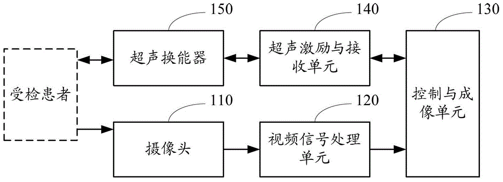
[0051] Such as figure 1 Shown is a schematic structural diagram of an endoscopic nasopharyngeal carcinoma ultrasound imaging device in an embodiment. The endoscopic ultrasound imaging device for nasopharyngeal carcinoma includes a camera 110 , a video signal processing unit 120 , a control and imaging unit 130 , an ultrasound excitation and reception unit 140 and an ultrasound transducer 150 connected in sequence.

[0052] Wherein, the camera 110 is used for collecting tissue optical images of the inner surface of the nasopharynx.

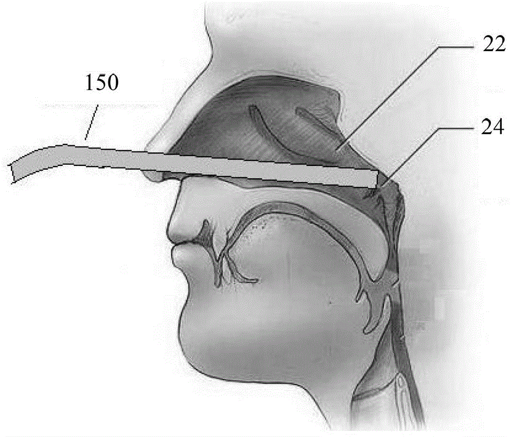
[0053] In this embodiment, the camera 110 is a high-definition camera. The camera 110 can extend into the nasopharynx through the nostrils to realize high-definition optical imaging of the inner surface of t...


PUM


Abstract

An endoscopic ultrasonic imaging device and method for nasopharynx cancer. The device comprises: a camera (110), used for acquiring an optical tissue image on the inner surface of a nasopharynx; a video signal processing unit (120), used for converting an optical tissue image signal into a video digital signal; a control and imaging unit (130), used for storing and displaying the digital signal and sending a control instruction; an ultrasonic excitation and receiving unit (140), used for generating a specific pulse signal according to an ultrasonic control instruction for ultrasonic excitation, processing an ultrasonic echo signal and then performing ultrasonic image display; and an ultrasonic transducer (150), used for generating an ultrasonic wave for scanning the interior of the nasopharynx according to the pulse signal, receiving the ultrasonic echo signal and converting the ultrasonic echo signal into an electric echo signal; the ultrasonic excitation and receiving unit (140) is also used for processing the electric echo signal; the control and imaging unit (130) is also used for performing ultrasonic imaging according to the processed electric echo signal. The detection accuracy is improved.

Description

technical field [0001] The invention relates to medical equipment, in particular to an endoscopic nasopharyngeal carcinoma ultrasonic imaging device. Background technique [0002] Nasopharyngeal carcinoma is a malignant tumor that originates in the nasopharynx. About 600,000 new patients with nasopharyngeal carcinoma are diagnosed every year in the world, ranking fifth in the incidence of malignant tumors. The early symptoms of nasopharyngeal carcinoma are not obvious, and the middle and advanced symptoms include bloody nose, bloody sputum, tinnitus, deafness, cervical lymphadenopathy, headache, facial numbness, diplopia, drooping eyelids, blindness, dysphagia, hoarseness, tongue deviation Wait. Most of the symptoms do not cause pain to the patient, and are easily confused with symptoms of other etiologies; usually, nasopharyngeal carcinoma occurs in the mucous membrane of the inner wall of the nasopharynx, which is relatively hidden, and it is difficult to accurately iden...

Claims


Application Information

Patent Timeline
Patent Type & Authority Patents(China)
IPC IPC(8): A61B8/08
CPCA61B8/12A61B1/233
Inventor 郑海荣邱维宝黎国锋梁长虹
Owner NAT INST OF ADVANCED MEDICAL DEVICES SHENZHEN
Why Eureka
Try Eureka