Automatic segmentation method of knee joint cartilage image

An automatic segmentation and image segmentation technology, which is applied in the field of image processing, can solve problems such as false edges, and achieve the effect of strong adaptability, good stability, and ideal segmentation effect

Inactive Publication Date: 2013-12-11
CHONGQING UNIV
View PDF7 Cites 31 Cited by
  • Summary
  • Abstract
  • Description
  • Claims
  • Application Information

AI Technical Summary

Problems solved by technology

[0007] In view of the above problems, the object of the present invention is to propose a method for automatic segmentation of knee articular cartilage images, using edge positioning based on SVM to solve the problem of false edges during edge detection, and simultaneously utilizing the self-adaptive region growing method for automatically selecting seed points for cartilage segmentation. Image segmentation to improve the accuracy of knee cartilage image segmentation

Method used


Image

  • Automatic segmentation method of knee joint cartilage image
  • Automatic segmentation method of knee joint cartilage image
  • Automatic segmentation method of knee joint cartilage image

Examples


Embodiment Construction

[0044] The present invention will be further described below in combination with specific embodiments and accompanying drawings. The specific embodiments described here are only used to explain the present invention, not to limit the present invention.

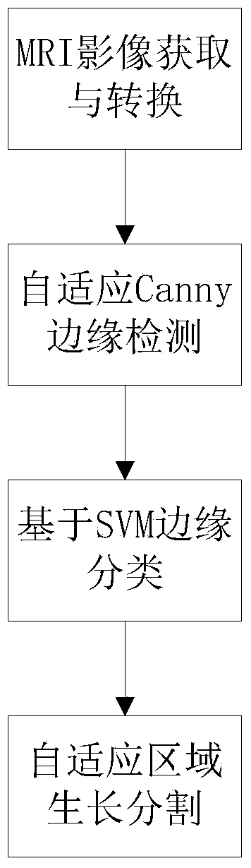
[0045] Such as figure 1 As shown, a method for automatic segmentation of knee articular cartilage images, including an edge location step based on SVM and an image segmentation step based on a region growing method, wherein:

[0046] The SVM-based edge localization steps include:

[0047] Step 11: Obtain the MRI image of the knee joint to be segmented and convert it into a grayscale image;

[0048]In this example, the MRI image of the right knee joint of a healthy adult male with no joint medical history is used as the research object. , resolution: 384×384). The images are numbered from 01 to 20 from outside to inside. Then convert the sequence original image in DICOM format into a grayscale image in jpg format, where th...


PUM


Abstract

The invention discloses an automatic segmentation method of a knee joint cartilage image. The method is characterized by comprising edge positioning based on SVM (Space Vector Modulation) and image segmentation based on a region growing method, wherein the step of edge positioning based on the SVM comprises acquisition and conversion of a knee joint MRI (Magnetic Resonance Imaging) image, adaptive Canny edge detection and cartilage edge classification based on the SVM; and in the step of image segmentation based on the region growing method, cartilage tissues are segmented by mainly adopting the improved region growing method capable of automatically selecting seed points. The method has the beneficial effects that the cartilage segmentation is performed on the knee joint MRI image; the precision positioning is realized by effectively combining the mode recognition with the edge detection; and the positioning complementation is sufficiently implemented in combination of the region growing method, so that the internal similar characteristics and the external difference characteristics of regions to be segmented are combined. Thus, the defects of result over-segmentation or inaccurate segmentation and the like of traditional segmentation method are effectively overcome.

Description

technical field [0001] The present invention relates to image processing technology, in particular to an automatic knee cartilage image segmentation method based on a support vector machine (Support Vector Machine, SVM) and a region growing method. Background technique [0002] The knee joint is the joint with the most complex structure and the most vulnerable joints in the human body. Its common diseases include arthritis, bone tumors, etc., and these diseases are often accompanied by the degeneration, destruction and morphological changes of articular cartilage. Early diagnosis is very important. As a non-invasive examination method, magnetic resonance imaging has become the main means to evaluate the morphology and function of cartilage. Segmenting articular cartilage by MRI imaging and then calculating its thickness, volume and other parameters can realize quantitative evaluation of cartilage and provide a strong diagnostic basis for clinical medicine, so as to take ear...

Claims


Application Information

Patent Timeline
Patent Type & Authority Applications(China)
IPC IPC(8): G06T7/00G06K9/62
Inventor 李勇明邹雪王品谢文宾吕洋何璇
Owner CHONGQING UNIV
Why Eureka
Try Eureka