
Method for extracting cells from night soil microscopic image

A technology of microscopic images and cells, applied in the field of biomedical image processing

Inactive Publication Date: 2014-02-26
HUAZHONG UNIV OF SCI & TECH
View PDF3 Cites 15 Cited by
  • Summary
  • Abstract
  • Description
  • Claims
  • Application Information

AI Technical Summary

Problems solved by technology

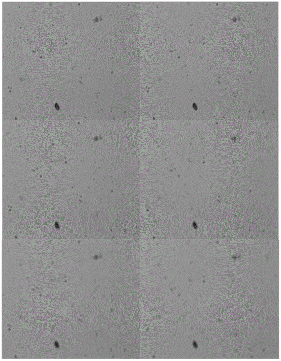
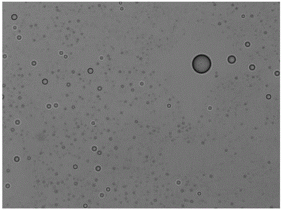
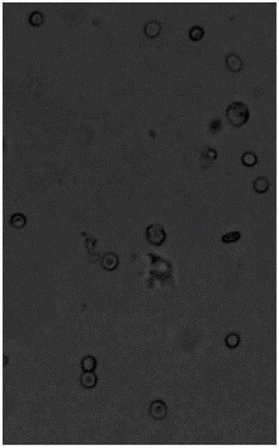
[0004] Aiming at the above defects or improvement needs of the prior art, the present invention provides a method for extracting cells from feces microscopic images, the purpose of which is to solve how to obtain image clarity through microscopic images to control the focus of the microscope, and how to extract cells from microscopic images Find the specific position of the cell and determine the technical problem of the cell type when the background has many impurities, the contrast between the cell and the background is low, and the cell edge is not obvious

Method used


Image

  • Method for extracting cells from night soil microscopic image
  • Method for extracting cells from night soil microscopic image
  • Method for extracting cells from night soil microscopic image

Examples


Embodiment Construction

[0056] In order to make the object, technical solution and advantages of the present invention clearer, the present invention will be further described in detail below in conjunction with the accompanying drawings and embodiments. It should be understood that the specific embodiments described here are only used to explain the present invention, not to limit the present invention. In addition, the technical features involved in the various embodiments of the present invention described below can be combined with each other as long as they do not constitute a conflict with each other.

[0057] The terms used in the present invention are firstly explained and described below.

[0058] Hough circle detection: that is, to detect objects that are approximately circular in the image through the Hough transform. The principle is as follows:

[0059] First, edge detection is performed on the image (the Canny operator is used here), and then the local gradient is considered for each ...


PUM


Abstract

The invention discloses a method for extracting cells from a night soil microscopic image. The method comprises steps that: (1), a cell microscopic image is shot by a camera and is stored into a computer, the cell microscopic image is read, an average gradient G is calculated, a focal length of a microscope is adjusted according to the G value, shooting and image reading are carried out continuously, till the most intelligible image IIN corresponding to the largest average gradient G is found out; fat balls are extracted from the most intelligible image IIN from the (1) step to acquire positions and the number of the fat balls of the IIN; and (3), cells are extracted from the most intelligible image IIN from the (1) step through an edge extraction and threshold segmentation method to acquire positions, types and the numbers of red blood cells and white blood cells of the IIN. Through the method, technical problems of how to acquire image sharpness through the microscopic image to control focusing of the microscope, how to find out specific positions of the cells under conditions of a plurality of background impurities, a low contrast ratio of the cells to the background and unconspicuous cell edges, and how to determine cell types are solved.

Description

technical field [0001] The invention belongs to the field of biomedical image processing, and more specifically relates to a method for extracting cells from feces microscopic images. Background technique [0002] In clinical pathology examination, cytomorphological examination is of great significance. As far as the blood cell automatic analysis system is concerned, its research focuses on: (1) segmenting cells from images; (2) extracting cell features; (3) identifying cell types. Among them, cell segmentation is the most critical technology, because the segmentation result will directly affect the quality of subsequent cell feature extraction and recognition. [0003] Most of the blood cell analyzers commonly used clinically analyze the cell components directly through the microscopic imaging images of human blood samples. Since the human blood cell fluid is translucent and contains less impurities, the background of such images is relatively clean, and the cells and The...

Claims


Application Information

Patent Timeline
IPC IPC(8): G06T7/00
Inventor 刘建国马显芳张桂贤
Owner HUAZHONG UNIV OF SCI & TECH
Learn More