
Lesion tissue growth monitoring method and system

A tissue growth and monitoring system technology, applied in the field of medical image processing, can solve the problems of low classification accuracy of benign and malignant tumors, inability to fully represent tumor elasticity information, etc., and achieve high accuracy, high classification accuracy, and strong robustness Effect

Active Publication Date: 2014-04-16
SHENZHEN INST OF ADVANCED TECH CHINESE ACAD OF SCI
View PDF6 Cites 26 Cited by
  • Summary
  • Abstract
  • Description
  • Claims
  • Application Information

AI Technical Summary

Problems solved by technology

[0009] Based on this, it is necessary to provide a method and system for monitoring the growth of lesion tissue in view of the problems that the above elastic characteristic values ​​still cannot fully represent the elastic information of the tumor and the classification accuracy of benign and malignant tumors is low.

Method used


Image

  • Lesion tissue growth monitoring method and system
  • Lesion tissue growth monitoring method and system
  • Lesion tissue growth monitoring method and system

Examples


Embodiment Construction

[0033] The specific implementations of the method and system for monitoring diseased tissue growth of the present invention will be described in detail below in conjunction with the accompanying drawings.

[0034] The lesion tissue growth monitoring method and system of the present invention can be applied to monitor the growth of various lesion tissues, for example, the evaluation of the elasticity of the liver, lung, thyroid, etc., the evaluation of the elasticity of the infiltration and growth characteristics of breast tumors, and can also be used for breast and breast tumors. Evaluation of the degree of elasticity of diseased areas on other parts of the human body.

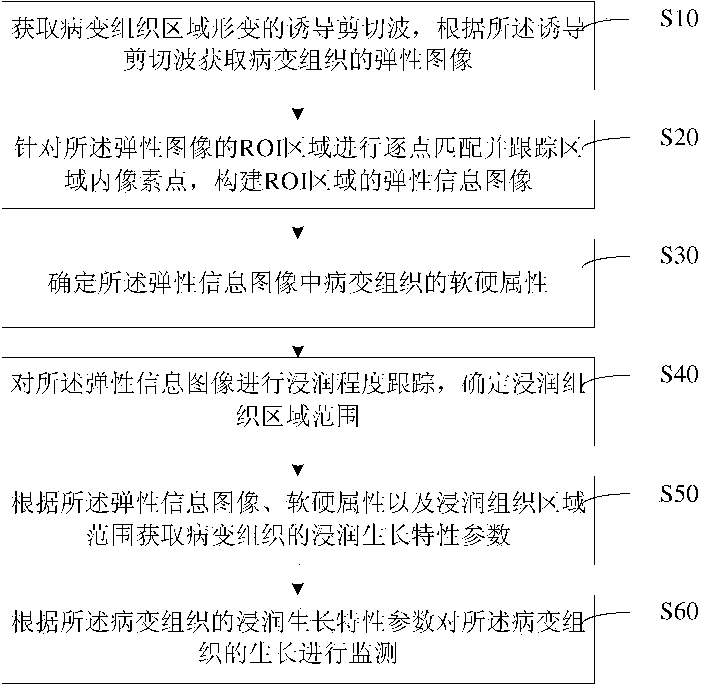
[0035] see figure 1 as shown, figure 1 It is a flowchart of a method for monitoring pathological tissue growth in an embodiment, which mainly includes the following steps:

[0036] Step S10, acquiring the induced shear wave of the deformation of the lesion tissue region, and acquiring the elastic image of th...


PUM


Abstract

The invention provides a lesion tissue growth monitoring method and system. The method includes the steps that inductive shear waves for deformation of a lesion tissue area are acquired, and an elastic image of lesion tissue is acquired according to the inductive shear waves; point-by-point matching is performed for an ROI area of the elastic image, pixel points in the area are tracked, and an elastic information image of the ROI area is constructed; the hardness attribute of the lesion tissue in the elastic information image is determined; the infiltration degree of the elastic information image is tracked, and the range of the infiltration tissue area is determined; the infiltration growth characteristic parameters of the lesion tissue are acquired according to the elastic information image, the hardness attribute and the infiltration tissue area range; the growth of the lesion tissue is monitored according to the infiltration growth characteristic parameters of the lesion tissue. Through the technical scheme, elastic information of the lesion tissue can be represented completely, the benign / malignance mass classification precision is high, the tumor growth monitoring accuracy is high, and an important basis is provided for pathological research on tumors and early diagnosis and treatment of tumors.

Description

technical field [0001] The invention relates to the technical field of medical image processing, in particular to a method and system for monitoring the growth of diseased tissue. Background technique [0002] Medical clinical research has shown that when a cell becomes lesioned, the biomechanical properties of itself and the surrounding normal tissue will change significantly. Using image information showing tissue elasticity parameters to quantitatively characterize, evaluate and monitor the growth characteristics of breast tumors is beneficial to breast preservation. The preoperative prediction of surgery, the definition of radiation therapy target area and the discrimination of benign and malignant tumors are of great significance. [0003] For example, for the monitoring of breast tumor growth characteristics based on elastic parameters, the current research work is mainly focused on how to construct elastic images, and there is a lack of prospective research on elastic...

Claims


Application Information

Patent Timeline
IPC IPC(8): A61B8/08
Inventor 肖杨张雪郑海荣牛丽丽王丛知
Owner SHENZHEN INST OF ADVANCED TECH CHINESE ACAD OF SCI
Learn More