A Calculation Method of Cone Cell Density Based on Image Recognition

A calculation method and image recognition technology, applied in the field of cone cell density calculation, can solve the problems of no automatic cone cell counting density, limited application, labor consumption, etc., achieve a high degree of automation, avoid result deviation, and improve detection efficiency. Effect

CN103745257BActive Publication Date: 2016-08-17SCHOOL OF OPHTHALMOLOGY & OPTOMETRY WENZHOU MEDICAL COLLEGE
3 Cites 1 Cited by

Patent Information

Authority / Receiving Office
CN · China
Patent Type
Patents(China)
Current Assignee / Owner
Publication Date
2016-08-17

Smart Images

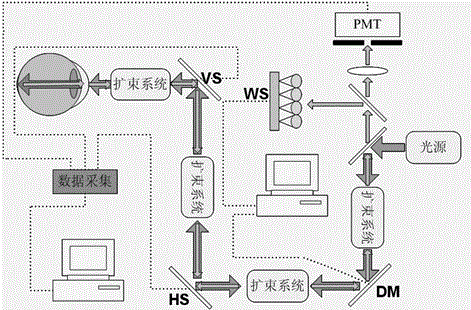
  • Figure 1
    Figure 1
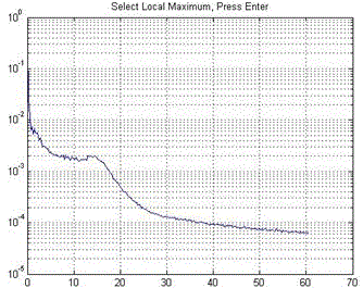
  • Figure 2
    Figure 2
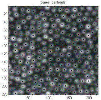
  • Figure 3
    Figure 3
Patent Text Reader

Abstract

The invention relates to a cone-cell density calculation method based on image identification. The method mainly solves the prior problem that density counting of cone cells is performed manually and an eye-ground adaptive confocal laser scanning ophthalmoscope is used to obtain a visual cone-cell image and after the response frequency of the cone cells is determined, low-pass filtering is used for denoising processing so as to establish a two-valued sequence. A local maximum value corresponding to a non-zero area is searched for so that the central position of the cone cells is found. After automatic identification of the visual cone cells in the image is completed, the number of the cone cells in the image is obtained with the assistance of manual correction and finally the density counting of the cone cells is achieved. When the function which adopts a computer to counting automatically is compared with a manual counting method, the automatic calculation method is accurate, high in statistical speed, high in automation degree, checkable in analysis result and improved in detection efficiency. At the same time, a manual correction function is added so that on the basis of software analysis, error identification can be removed manually and missed cone cells can be added and thus caused result deviation is prevented.
Need to check novelty before this filing date? Find Prior Art

Description

technical field

[0001] The invention belongs to the fields of fundus microscopic imaging and medical detection. Specifically, the invention relates to a method for calculating cone cell density based on image recognition. Background technique

[0002] There are three levels of neurons in the retina, including cones, rods, bipolar cells, and ganglion cells. Cone cells and rod cells belong to the first-level neurons. They sense external light and image information and transmit them to the visual center through bipolar cells and ganglion cells. Among them, the cone cells are mainly responsible for brightening vision, distinguishing fine shapes and color vision, and are mainly distributed in the macular area of ​​the retina (the center of the macula only has cone cells), which is the anatomical and physiological basis for the formation of visual acuity, that is, vision. And in some fundus diseases such as retinitis pigmentosa and cone-rod dystrophy, there will be changes in con...

Examples

Embodiment Construction

[0028] Embodiments of the present invention will be further described below in conjunction with accompanying drawings:

[0029] Such as Figure 1-Figure 4 As shown, the present invention discloses a method for calculating the density of cone cells based on image processing. The method utilizes a fundus adaptive confocal laser scanning ophthalmoscope to image fundus cone cells, and based on computer vision, the images are identified, analyzed and counted, and then counted. Cones, to analyze cone density. Said method comprises the following steps:

[0030] (1) Obtain pictures of retinal cones.

[0031] Turn on the power, enter the subject's personal information, and the subject sits in front of the fundus adaptive confocal laser scanning ophthalmoscope, with the lower jaw placed on the jaw rest.

[0032] Instruct the subject to stare at the red fixation light in the instrument, adjust the focus to make the cone cell picture of the fundus scanned and captured the clearest, and...