Handheld type fundus camera

A hand-held, camera technology, applied in the field of diagnosis and disease, can solve the problems of inconvenient for special patients, large size, and the machine does not have the image storage function, etc., and achieve the effect of simplifying the lighting device

Inactive Publication Date: 2014-05-28
SUZHOU MICROCLEAR MEDICAL INSTR
View PDF10 Cites 22 Cited by
  • Summary
  • Abstract
  • Description
  • Claims
  • Application Information

AI Technical Summary

Problems solved by technology

[0005] Traditional desktop fundus cameras include complex lighting systems and observation systems, which are huge in size and complex in system structure; some instruments require special software installed on a computer before they can be used, and the machines themselves do not have image storage functions, and some even cannot be separated from the computer Work independently
If pictures are needed, the patient needs to go to the instrument to take fundus pictures, whi

Method used


Image

  • Handheld type fundus camera
  • Handheld type fundus camera
  • Handheld type fundus camera

Examples


Embodiment Construction

[0039] The preferred embodiments of the present invention will be described in detail below in conjunction with the accompanying drawings, so that the advantages and features of the present invention can be more easily understood by those skilled in the art, so as to define the protection scope of the present invention more clearly.

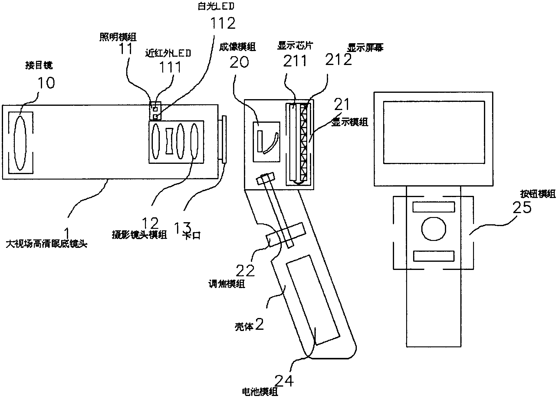
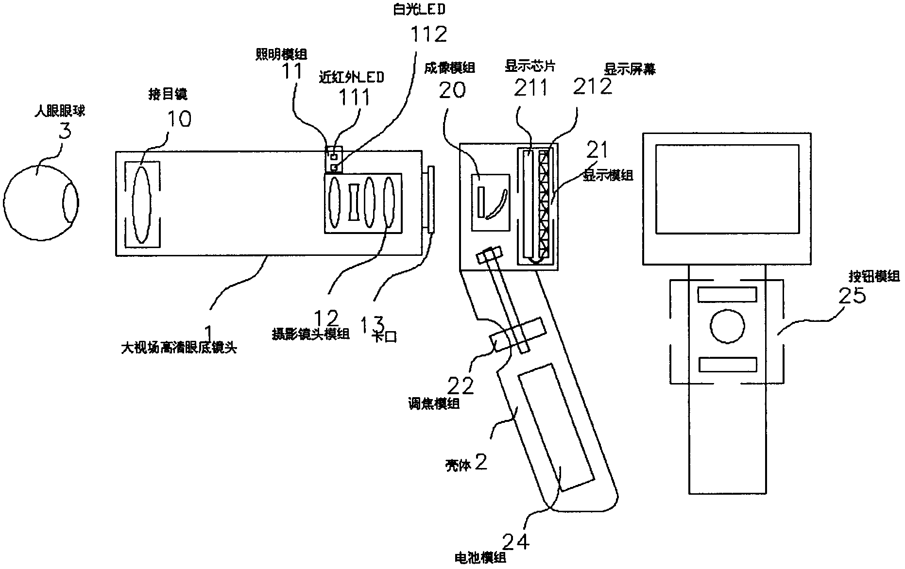
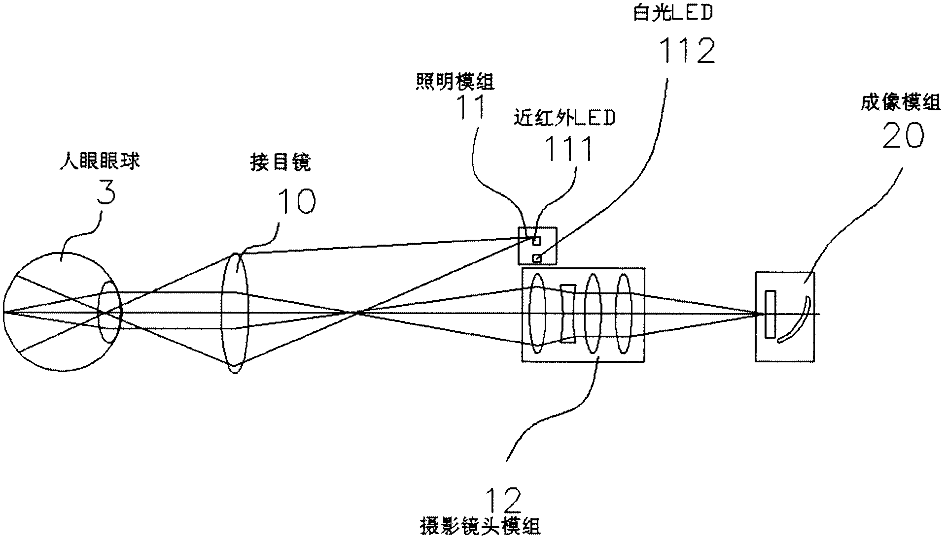
[0040] see figure 1 , the embodiment of the present invention includes: a hand-held fundus camera, which includes: 1. a large field of view high-definition fundus lens; 2. a housing; 3. an eyeball; 10. an eyepiece; 112. White light LED; 12. Photographic lens module; 13 Bayonet 20. Imaging module; 22. Display module, 201. CCD\COMS; 211. Display chip; 212. Display screen; 22. Focusing module; 23. Button module, 24. Battery module

[0041] The large-field-of-view high-definition fundus lens 1 includes an eyepiece 10 , an illumination group 11 , and a photographic lens module 12 . The lighting group 11 is located at the upper end of the photographi...


PUM


Abstract

The invention discloses a handheld type fundus camera used for fundus examination on human eyes medically. Human-eye fundus images are obtained through the optical imaging method in order to judge pathologies and focuses of fundus diseases. According to the handheld type fundus camera, a near-infrared LED and a white light LED are adopted as lighting light sources, optical lenses are used for collecting the fundus images, a CCD or COMS is used for receiving signals, and the human-eye fundus images are displayed on a screen through image transmission and processing. The handheld type fundus camera is simple in structure and capable of being carried around; a manual focusing mode can be adopted, imaging is clear and real-time image transmission can be achieved.

Description

technical field [0001] The present invention relates to a hand-held fundus camera, in particular to a large field of view fundus camera based on LED lighting and CCD\COMS imaging. Near-infrared and white LEDs are used as illumination sources to collect human fundus images through optical lenses. CCD or CCD COMS receives the signal and displays the fundus image of the human eye on the screen through hardware and software image processing. [0002] The invention belongs to the field of medical technology, and takes images of the fundus of the human eye, which are used for the screening of fundus diseases and assisting the diagnosis and condition judgment of other systemic diseases in the whole body, and involve image acquisition, image processing, network transmission and other technologies. Background technique [0003] LED light source is a new lighting method that has appeared in recent years. Compared with traditional light sources, LED has the advantages of long life, ene...

Claims


Application Information

Patent Timeline
IPC IPC(8): A61B3/14A61B3/12
Inventor 邱海洋张锐进李超宏
Owner SUZHOU MICROCLEAR MEDICAL INSTR
Why Eureka
Try Eureka