Contrast-enhanced ultrasound imaging method and contrast-enhanced ultrasonic imaging device

A technology of contrast-enhanced ultrasound and imaging methods, which can be used in measuring devices, ultrasonic/sonic/infrasonic diagnosis, and sound wave diagnosis, and can solve the problems of decreased contrast between blood vessels and tissues, long projection periods, and large cumulative errors.

Active Publication Date: 2014-06-25
SHENZHEN MINDRAY BIO MEDICAL ELECTRONICS CO LTD +1
View PDF5 Cites 6 Cited by
  • Summary
  • Abstract
  • Description
  • Claims
  • Application Information

AI Technical Summary

Problems solved by technology

On the other hand, the conventional MIP imaging technology is a cumulative and overlapping technique of contrast images. The longer the proje

Method used


Image

  • Contrast-enhanced ultrasound imaging method and contrast-enhanced ultrasonic imaging device
  • Contrast-enhanced ultrasound imaging method and contrast-enhanced ultrasonic imaging device
  • Contrast-enhanced ultrasound imaging method and contrast-enhanced ultrasonic imaging device

Examples


Embodiment 1

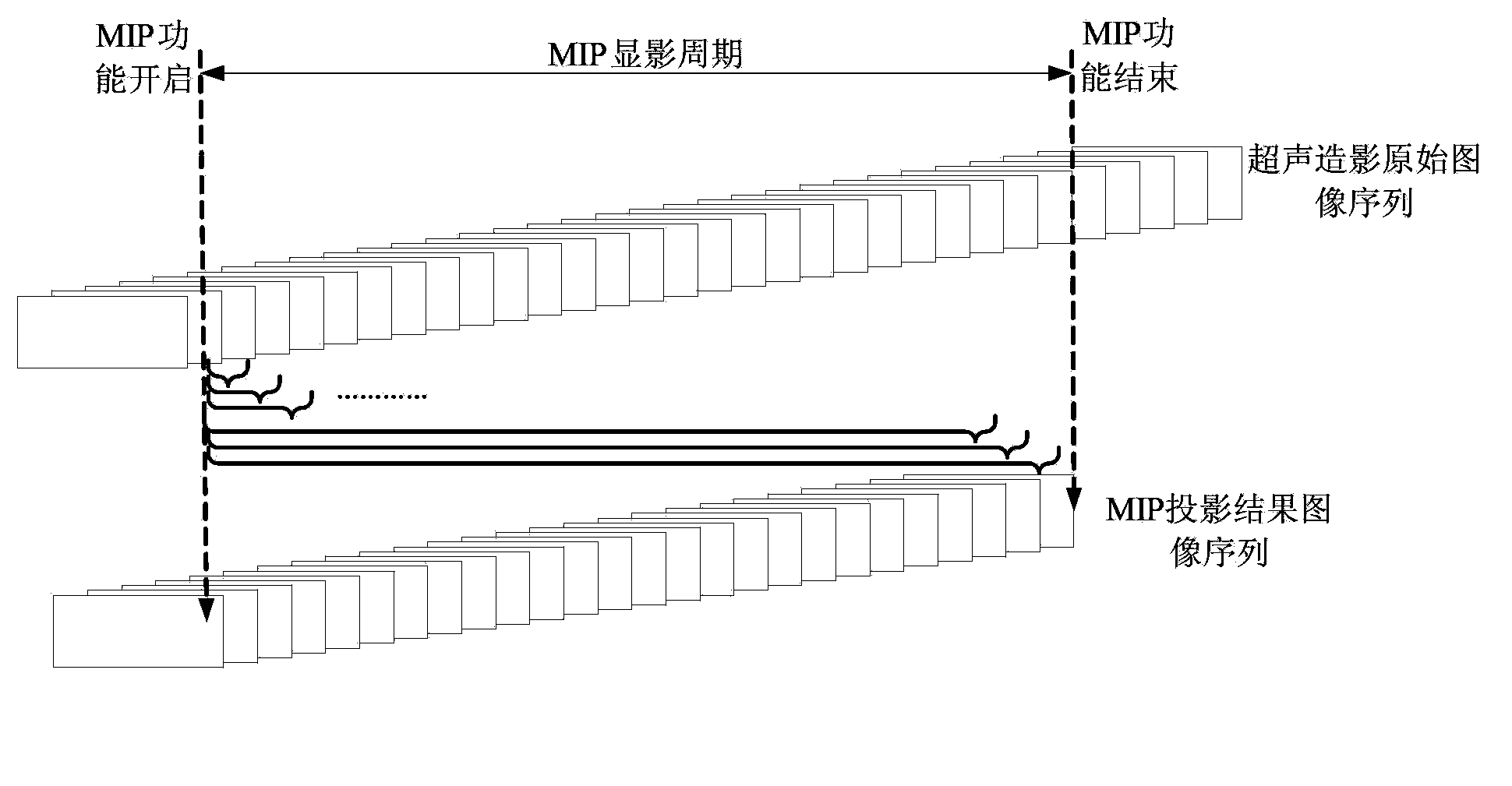
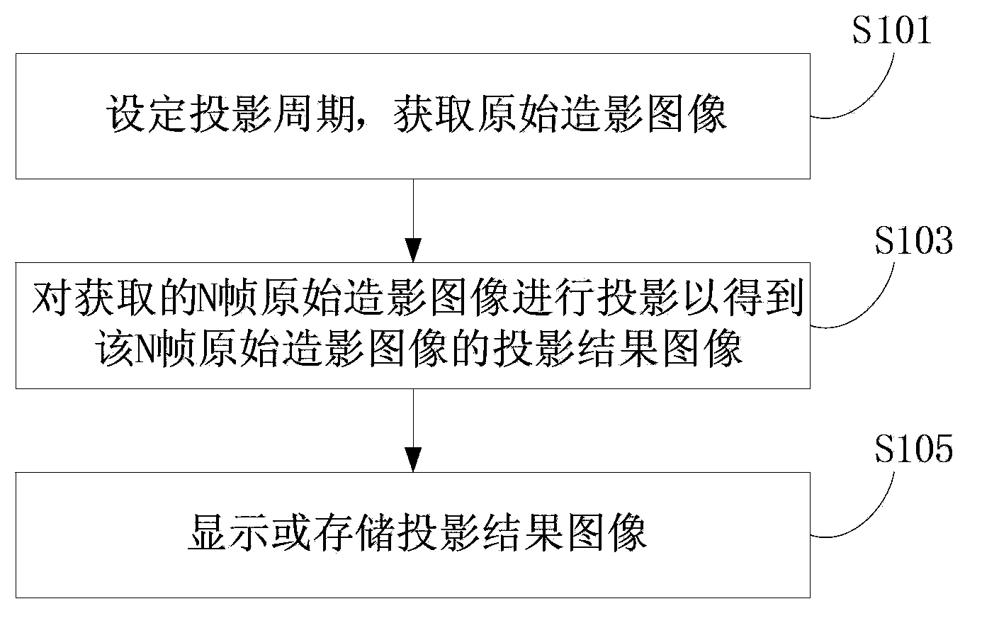
[0032] like image 3 As shown, the contrast-enhanced ultrasound imaging method of this embodiment includes the following steps S101 to S105:

[0033] The initial step S101 is to set the projection period and acquire N frames of original contrast images, where N is the total number of frames in the imaging period. The specific method of obtaining the original contrast image can refer to the commonly used ultrasonic imaging method. For example, firstly, the probe sends ultrasonic pulses to the object to be detected, and the probe responds and receives the ultrasonic echo signal returned by the object to be detected. Then, the system responds to the received ultrasonic echo signal. The wave signal is processed to output the original image at multiple times. Original images include B-mode images (ie, tissue images), contrast images, and the like. Here, the specific structure of the probe, its transmitting and receiving process, and signal processing can be realized by correspond...

Embodiment 2

[0065] During the process of ultrasound imaging, the scanned image inevitably moves, which affects the effect of contrast-enhanced ultrasound imaging. Image motion can be caused by tissue movement with breathing or heartbeat, or by movement of the ultrasound probe. Therefore, it is necessary to perform motion matching on multi-frame scanned images.

[0066] like Image 6 As shown, the processing of the contrast-enhanced ultrasound imaging method in this embodiment includes the following steps S201-S205:

[0067] Initial step S201, which is similar to step S101 in Embodiment 1, and will not be repeated here.

[0068] In the motion registration step S202, motion registration is performed on the N frames of original contrast images obtained in step S201.

[0069] In a specific ultrasound imaging system / device, the activation or deactivation of the step of motion registration can be realized in various ways, for example, the system is preset to start motion registration to perf...


PUM


Abstract

Provided are an ultrasound contrast imaging method and apparatus. The method comprises: an initial step (S101), acquiring N frames of original contrast images; a projection imaging step (S103), projecting on the N frames of original contrast images to obtain a projection result image of the N frames of original contrast images, wherein for an nth frame of original contrast image, any frame of an nth group of original contrast images is used as a projection template for projecting on the nth group of original contrast images to acquire a projection result image of the nth frame of original contrast image, the nth group of original contrast images is within a projection period and comprises several frames of original contrast images of the nth frame of original contrast image, and the projection period is smaller than an imaging period; and a showing and storing step (S105), showing or storing the projection result image of the N frames of original contrast images. In the method, the projection period is a fixed value, accumulated errors caused by a projection process are only related to the projection period, and cannot increase as the imaging period increases, so that images continuously projected onto a template can better reflect spatial information, such as a filling and regression state of a micro vessel, of test subject in a current projection period.

Description

technical field [0001] The present application relates to the field of ultrasound imaging, in particular to an ultrasound contrast imaging method and device. Background technique [0002] In the medical ultrasonic imaging system, the visible ultrasonic image of human tissue is obtained by receiving and processing the echo carrying the characteristic information of human tissue by using the reflection of ultrasonic wave at the interface of human tissue; however, due to the existence of reverberation and the limitation of resolution , and the influence of less blood flow and low blood flow velocity in the tiny blood vessels in the human tissue, it is difficult to visualize small blood vessels in color Doppler, and the display is sometimes blurred or even impossible to display. At present, contrast-enhanced ultrasound technology is generally used, which uses contrast agents to enhance backscattering, so that blood flow can be clearly displayed, thereby assisting doctors to achi...

Claims


Application Information

Patent Timeline
IPC IPC(8): A61B8/00
CPCG01S7/52034A61B8/00A61B8/481A61B8/5269A61B8/13A61B8/0891G01S15/8993G01S7/52039A61B8/483A61B8/5207A61B8/5276A61B8/461
Inventor 朱子俨桑茂栋董永强
Owner SHENZHEN MINDRAY BIO MEDICAL ELECTRONICS CO LTD
Why Eureka
Try Eureka