A 3D Cardiac Magnetic Resonance Imaging Method Based on Tensor Decomposition Sparse Constraints

A technology of magnetic resonance imaging and tensor decomposition, which is used in diagnostic recording/measurement, medical science, sensors, etc., which can solve the problem of reducing the number of imaging gradient coding steps, increasing the amount of calculation, and not considering the layer and layer sparseness of the three-dimensional cardiac magnetic resonance image. issues of sex

Active Publication Date: 2016-02-03
SUZHOU LONWIN MEDICAL SYST
View PDF3 Cites 0 Cited by
  • Summary
  • Abstract
  • Description
  • Claims
  • Application Information

AI Technical Summary

Problems solved by technology

[0004] At present, there are two main methods of MRI image reconstruction: one is multi-coil parallel imaging technology, which mainly uses the spatial sensitivity difference of a single receiving coil in the phased array coil to encode spatial information and reduce the gradient necessary for imaging. The number of encoding steps, which uses a multi-coil array to simultaneously collect signals, allows under-sampling of K space to reduce the number of phase encoding steps, while maintaining the same spatial resolution of the image, it can greatly shorten the scanning time and improve the imaging speed; but Multi-coil parallel imaging technology involves the estimation of multi-coil sensitivity distribution, which needs to increase the amount of calculation
[0005] Another MRI reconstruction method based on compressive sensing theory. Due to the sparse nature of magnetic resonance images, compressive sensing theory can be used to reconstruct images from randomly undersampled k-space data, reducing sampling data and improving imaging speed. Commonly used sparse transformation methods such as singular value decomposition, discrete wavelet transform, and discrete cosine transform only consider the sparse representation of single-layer cardiac magnetic resonance images, but do not consider the sparsity between layers of three-dimensional cardiac magnetic resonance images

Method used


Image

  • A 3D Cardiac Magnetic Resonance Imaging Method Based on Tensor Decomposition Sparse Constraints
  • A 3D Cardiac Magnetic Resonance Imaging Method Based on Tensor Decomposition Sparse Constraints
  • A 3D Cardiac Magnetic Resonance Imaging Method Based on Tensor Decomposition Sparse Constraints

Examples


Embodiment Construction

[0043] In order to describe the present invention more specifically, the technical solutions of the present invention will be described in detail below in conjunction with the accompanying drawings and specific embodiments.

[0044] Such as Figure 4 As shown, the three-dimensional cardiac magnetic resonance imaging method based on the tensor decomposition sparse constraint of the present embodiment, the specific implementation steps are as follows:

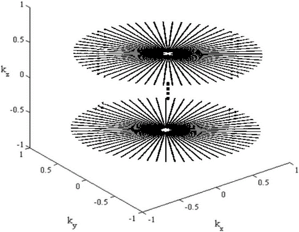
[0045](1) The undersampling of K-space data is realized by using three-dimensional radial sampling trajectories. A three-dimensional radial sampling trajectory, which contains N z sampling levels, each level contains N p projection lines, and each projection line contains N s sample points. Each sampling level is surrounded by the first level k z The axis is rotated for a certain amount, such as figure 1 As shown, the three-dimensional coordinates of the sampling points of the three-dimensional radial sampling trajectory (G...


PUM


Abstract

The invention discloses a three-dimensional heart magnetic resonance imaging method based on tensor composition sparse bound. According to the method, undersampling of whole heart three-dimensional K spatial data is achieved through a three-dimensional radial sampling track; sparse representation of a magnetic resonance image is achieved through high-order tensor decomposition, optimal sparse representation of whole heart three-dimensional magnetic resonance data is achieved, and magnetic resonance imaging accuracy is improved; L1 norms of tensor decomposition sparse data and composite regularization terms are combined to serve as bound terms, reconstruction of the undersampled three-dimensional heart magnetic resonance image is achieved through a rapid composite splitting algorithm. By the adoption of the method, magnetic resonance imaging scanning time is effectively shortened, the heart magnetic resonance imaging speed is increased, motion artifacts generated in the heart detection process can be conveniently eliminated, and three-dimensional heart magnetic resonance imaging accuracy is improved.

Description

technical field [0001] The invention belongs to the technical field of magnetic resonance imaging, and in particular relates to a three-dimensional cardiac magnetic resonance imaging method based on tensor decomposition sparse constraints. Background technique [0002] Cardiovascular disease is the leading cause of sudden cardiac arrest. At present, the incidence and mortality of cardiovascular diseases remain high, which increases the burden of cardiovascular disease prevention and treatment and becomes an important public health problem. It is urgent to strengthen the prevention and treatment of cardiovascular diseases. Cardiac Magnetic Resonance Imaging (CMR) is a technology that uses the principle of nuclear magnetic resonance to perform tomographic imaging of the human heart. It can accurately reflect the anatomical structure, morphological function, blood flow characteristics and myocardial activity of the heart. main tool. Cardiac magnetic resonance imaging has good...

Claims


Application Information

Patent Timeline
Patent Type & Authority Patents(China)
IPC IPC(8): A61B5/055
Inventor 蒋明峰汪亚明黄文清冯杰郑俊褒
Owner SUZHOU LONWIN MEDICAL SYST
Why Eureka
Try Eureka