Geometric calibration method for SPECT (single photon emission computed tomography) system

A technology of single-photon emission and tomographic imaging, which is applied in computerized tomography scanners, echo tomography, etc., can solve problems such as complex scanning orbits and data fitting methods, difficult operations, and difficulties, and achieve the effect of improving imaging quality

Active Publication Date: 2014-12-03
BEIJING NOVEL MEDICAL EQUIP LTD
View PDF4 Cites 26 Cited by
  • Summary
  • Abstract
  • Description
  • Claims
  • Application Information

AI Technical Summary

Problems solved by technology

[0005] The traditional method needs to prepare a smaller-sized point source, which has certain difficulties. At th

Method used


Image

  • Geometric calibration method for SPECT (single photon emission computed tomography) system
  • Geometric calibration method for SPECT (single photon emission computed tomography) system
  • Geometric calibration method for SPECT (single photon emission computed tomography) system

Examples


Embodiment Construction

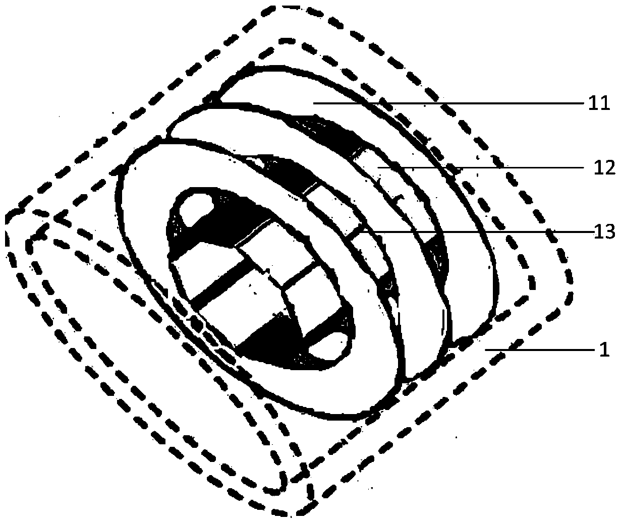
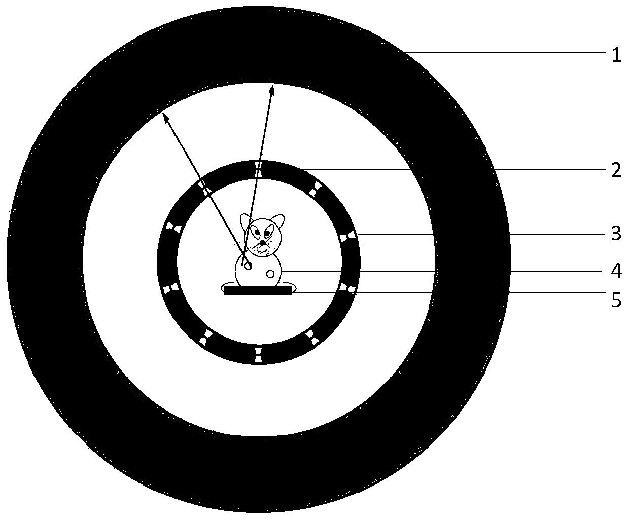
[0034] Embodiments of the present invention are described in detail below, examples of which are shown in the drawings, wherein the same or similar reference numerals designate the same or similar elements or elements having the same or similar functions throughout. The embodiments described below by referring to the figures are exemplary only for explaining the present invention and should not be construed as limiting the present invention.

[0035] In describing the present invention, it should be understood that the terms "center", "longitudinal", "transverse", "upper", "lower", "front", "rear", "left", "right", " The orientations or positional relationships indicated by "vertical", "horizontal", "top", "bottom", "inner" and "outer" are based on the orientations or positional relationships shown in the drawings, and are only for the convenience of describing the present invention and Simplified descriptions, rather than indicating or implying that the device or element refe...


PUM


Abstract

The invention discloses geometric calibration method for a SPECT (single photon emission computed tomography) system. The method comprises the steps as follows: a collimator is measured to obtain geometric parameters of a pinhole or a slot in the collimator; the collimator and a PET (position emission tomography) detector are assembled to form the SPECT system; the collimator is controlled to move, and the PET detector is used for measuring multi-group background coincidence events when the collimator moves to a plurality of target positions; and position coordinates of the collimator in the SPECT system are obtained according to the multi-group background coincidence events. According to the method, the geometric parameters of the SPECT system can be accurately calibrated, accurate geometric parameters are provided for image reconstruction of tomography, and accordingly, the imaging quality of the SPECT system is improved.

Description

technical field [0001] The invention relates to the technical field of emission tomography, in particular to a geometric calibration method for a single-photon emission tomography system. Background technique [0002] Emission tomography is a non-invasive nuclear medicine imaging method. Its principle is to use radionuclide-labeled biologically active substances to participate in the physiological metabolism of organisms, and to indirectly observe the metabolism of biologically active substances by measuring the distribution of radionuclides. and three-dimensional distribution. Emission tomography includes SPECT (Single Photon Emission Computed Tomography) and PET (Positron Emission Tomography), both of which are functional imaging techniques and have been widely used in pre-clinical drug research and clinical disease diagnosis. Compared with PET, SPECT has the advantages of high resolution and more kinds of labeled drugs. [0003] A current construction method of a SPECT ...

Claims


Application Information

Patent Timeline
IPC IPC(8): A61B6/03
Inventor 魏清阳刘亚强王石马天予江年铭刘迈
Owner BEIJING NOVEL MEDICAL EQUIP LTD
Why Eureka
Try Eureka