Eureka AIR delivers breakthrough ideas for toughest innovation challenges, trusted by R&D personnel around the world.

A fixed observation device for live imaging of animal spinal cord

A living imaging and observation device technology, which is applied in the fields of animal restraint equipment, medical science, sensors, etc., can solve the problems of increasing inflammatory response, harming the animal's systemic function, and difficulty in postoperative imaging fixation, etc., and achieves a wide range of applications.

Inactive Publication Date: 2016-11-09
GENERAL HOSPITAL OF PLA
View PDF6 Cites 2 Cited by
  • Summary
  • Abstract
  • Description
  • Claims
  • Application Information

AI Technical Summary

Problems solved by technology

However, this process cannot last for a long time. Prolonged anesthesia and repeated operations will not only harm the animal's systemic function, but also increase the risk of infection, increase the inflammatory response, cause tissue damage, and cause more pain to the animal. Not conducive to further observation and experimental research
Moreover, there is currently no uniform standard for surgical fixation and postoperative observation of external fixators, resulting in complicated surgical operations and difficulties in postoperative imaging and fixation.

Method used


Image

  • A fixed observation device for live imaging of animal spinal cord
  • A fixed observation device for live imaging of animal spinal cord
  • A fixed observation device for live imaging of animal spinal cord

Examples


Embodiment Construction

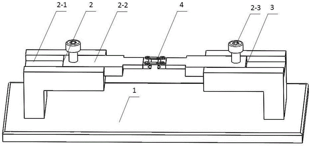
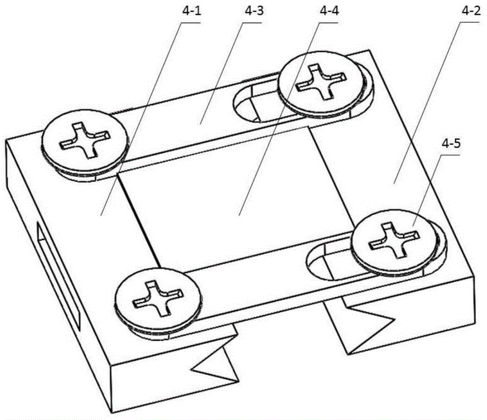
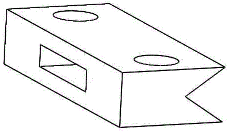
[0037] See Figure 1-Figure 4 , the technical solution of the present invention is: a kind of animal spinal cord live imaging fixed observation device, it is made up of base plate 1, left side movement adjustment device 2, right side movement adjustment device 3, implantable observation window 4; The positional connection relationship is: the left mobile adjustment device 2 and the right mobile adjustment device 3 are fixed on the bottom plate 1 through screws, and the implanted observation window 4 is connected with the left mobile adjustment device 2 and the right mobile adjustment device through the upper rectangular hole. The rectangular protrusions of the device 3 are fitted and positioned.

[0038] The installation 1 is a rectangular plate, and two mounting threaded holes are respectively arranged on the left and right sides for fixing the left side movement adjustment device 2 and the right side movement adjustment device 3 .

[0039] Described left side movement regul...


PUM


Abstract

A fixing and observing device for living imaging of animal spinal cords comprises a bottom plate (1), a left movement adjusting device (2), a right movement adjusting device (3) and an embedded observation window (4). The left movement adjusting device (2) and the right movement adjusting device (3) are fixedly connected to the bottom plate (1) through screws. The embedded observation window (4) is in insertion connection with rectangular protrusions of horizontal positioning sliders on the left movement adjusting device (2) and the right movement adjusting device (3) through rectangular holes in V-shaped frames to be positioned. By the fixing and observing device, operations for installing the embedded observation window can be facilitated to improve stability of the spinal cords, and dynamic observation of post-operation two-photon spinal-cord imaging can be also facilitated. The horizontal positioning sliders on the left movement adjusting device and the right movement adjusting device can guarantee horizontal stable internal fixation states for implant operations and can further provide stable external fixation support during post-operation observation.

Description

Technical field: [0001] The invention relates to a fixed observation device for living animal spinal cord imaging, which is mostly used in experimental research in the field of neurobiology, can perform implanted observation window installation operations on animals, and can perform long-term dynamic observation of spinal cord nerve tissue and its components in vivo. Changes in blood vessels are of great value in the study of the central nervous system, especially the physiological and pathological changes of spinal cord injury, as well as the regeneration and repair of nerves after spinal cord injury. Background technique: [0002] In the experimental research in the field of neurobiology, the traditional method of in vivo imaging of the animal spinal cord is to excise a segment of the lamina to expose the spinal cord, use the whole body suspension method or local clamping to stabilize the spine, and then use the fluorescence microscope or two-photon Microscopy for in vivo ...

Claims


Application Information

Patent Timeline
Patent Type & Authority Patents(China)
IPC IPC(8): A61D3/00
CPCA61B5/0062A61B5/0082A61B5/407A61B5/48A61D3/00
Inventor 张逸凌唐佩福张立海季欣然陈超张浩张金辉
Owner GENERAL HOSPITAL OF PLA
Why Eureka
Eureka Blog
Learn More