Cone beam CT (computed tomography) area-of-interest scanning method based on visualization
A technology of region of interest and scanning method, which is applied in the field of secondary scanning of the region of interest of cone beam CT to achieve accurate results
- Summary
- Abstract
- Description
- Claims
- Application Information
AI Technical Summary
Problems solved by technology
Method used
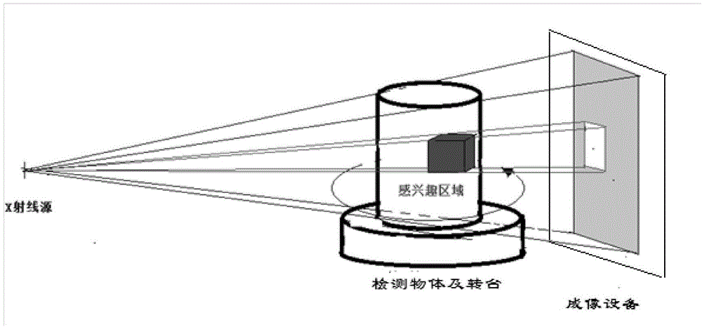
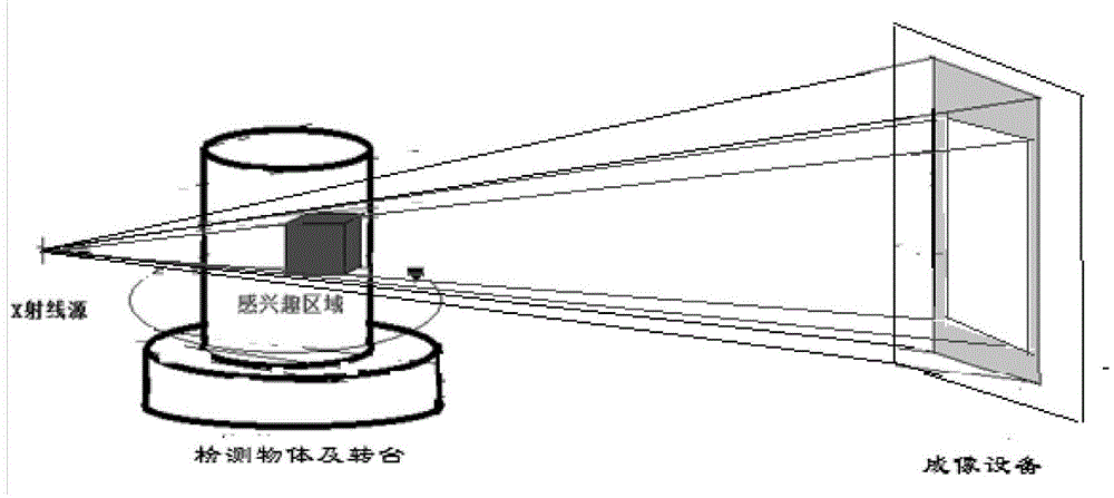
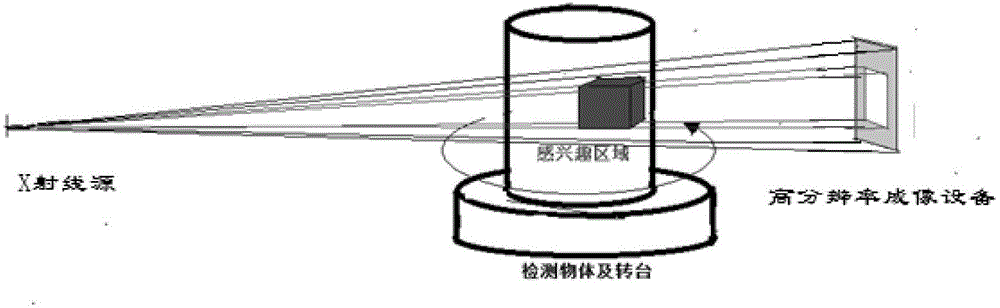
Image

Examples
Embodiment 1
[0035] Example 1 Reconstruction of the mouse kidney region of interest
[0036] Such as Figure 4 and Figure 5 , first open the folder where the picture is located, and load all the data into the memory. Then obtain the region of interest according to the cone beam CT secondary scanning method of the present invention:
[0037] The first step: the overall initial scan to obtain the overall data of the whole mouse, and perform three-dimensional reconstruction through the visualization method.
[0038] Step 2: Enable the combined display function of the multi-plane reconstruction effect and the 3D reconstruction effect.
[0039] Step 3: Three-dimensional cropping to the preliminary area. Since the region of interest is in the kidney of the mouse, it is initially cropped to the corresponding position
[0040] Step 4: Move the multi-plane reconstruction plane data, determine the exact position and move the cropping frame to the specified position, and determine the precise ra...
PUM
 Abstract
Description
Claims
Application Information
 - R&D
- Intellectual Property
- Life Sciences
- Materials
- Tech Scout
- Unparalleled Data Quality
- Higher Quality Content
- 60% Fewer Hallucinations
Browse by: Latest US Patents, China's latest patents, Technical Efficacy Thesaurus, Application Domain, Technology Topic, Popular Technical Reports.
© 2025 PatSnap. All rights reserved.Legal|Privacy policy|Modern Slavery Act Transparency Statement|Sitemap|About US| Contact US: help@patsnap.com