X-ray chest radiography lung segmentation method and device

A lung and chest X-ray technology, applied in the field of biomedical images, can solve the problem of not giving the outline of the lungs

Active Publication Date: 2015-03-18
SHENZHEN INST OF ADVANCED TECH
View PDF3 Cites 29 Cited by
  • Summary
  • Abstract
  • Description
  • Claims
  • Application Information

AI Technical Summary

Problems solved by technology

However, the defect of the rule-based boundary detection method is that only the main lung boundary segments are given, and the complete lung contour is not given.

Method used


Image

  • X-ray chest radiography lung segmentation method and device
  • X-ray chest radiography lung segmentation method and device
  • X-ray chest radiography lung segmentation method and device

Examples


Embodiment Construction

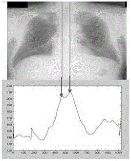
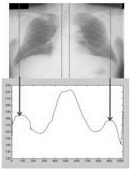
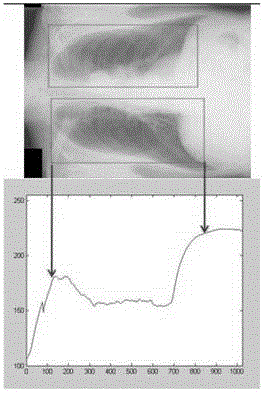
[0038] An embodiment of the present invention provides a method for lung segmentation in an X-ray chest film, including: S101, acquiring two rectangular regions respectively surrounding the left lung image and the right lung image in the X-ray chest film through horizontal and vertical projection; S102, Initialize the lungs in the two rectangular areas to obtain the initial shape of the lungs; S103, according to the weighted grayscale local texture model, find each feature point (x i ,y i ) of the best matching point (x’ i ,y' i ), remember dI X =(dx 1 , d y 1 , d x 2 , d y 2 ,..., d x i , d y i ,..., d x n , d y n ) T , the d x i =x' i -x i , the d y i =y' i -y i , the i=1, 2,..., n, the lung image includes the lung shape in the adjustment stage and the initial shape of the lung; S104, by adjusting the attitude parameter and shape parameter b, the current shape of the lung is I Xc as close as possible to I X +dI X , the adjustment result of the shape para...


PUM


Abstract

The invention discloses an X-ray chest radiography lung segmentation method so as to carry out accurate segmentation on lungs in an X-ray chest radiography. The method includes the following steps: S101: through horizontal and vertical projection, obtaining two rectangular areas which respectively surround left and right lung images in the X-ray chest radiography; S102: initializing the lungs in the two rectangular areas so as to obtain the initial shapes of the lungs; S103: according to a weighted grey local texture model, searching for optimal matching points of characteristic points in the lung images; S104: through adjustment of attitude parameters and a shape parameter b, enabling current shapes I<Xc> of the lungs to approximate I<X>+dI<X> in the largest degree; and repeating S103 and S104 until the change quantities are smaller than preset thresholds when obtained lung shapes are compared with lung shapes obtained through a previous adjustment next to a current adjustment. The method and device are capable of obtaining better initialization lung shapes and do not cause an over-segmentation phenomenon in a follow-up adjustment process and are capable of carrying out accurate segmentation on lung areas in the X-ray chest radiography under the restraint of an active shape model.

Description

technical field [0001] The invention relates to the field of biomedical images, in particular to a lung segmentation method and device for an X-ray chest film. Background technique [0002] Chest X-ray examination is still the most important means of initial detection of lung diseases. In clinical applications, chest X-ray images are widely used in the diagnosis of lung diseases. The first step in many chest X-ray image analysis applications is to mark the boundaries of the lungs in the image so that the computer can carry out the next step of processing. Therefore, the lung Segmentation is very important. Firstly, because the area of ​​the lungs accounts for 1 / 3 of the entire X-ray chest film, if the area of ​​interest in the lungs is extracted during diagnosis, the accuracy of the diagnosis will be greatly improved; secondly, after the lungs are segmented, other Processing will greatly reduce the interference of other irrelevant tissues and improve the speed of diagnosis...

Claims


Application Information

Patent Timeline
Patent Type & Authority Applications(China)
IPC IPC(8): G06T7/00A61B6/00
CPCA61B6/5229G06T7/11G06T2207/10116G06T2207/30061
Inventor 胡庆茂吴刚
Owner SHENZHEN INST OF ADVANCED TECH
Why Eureka
Try Eureka