Eureka AIR delivers breakthrough ideas for toughest innovation challenges, trusted by R&D personnel around the world.

X-ray computed tomography apparatus and information processing apparatus

A tomography, X-ray technology used in computing, image data processing, computed tomography, etc.

Active Publication Date: 2019-09-20
TOSHIBA MEDICAL SYST CORP
View PDF9 Cites 0 Cited by
  • Summary
  • Abstract
  • Description
  • Claims
  • Application Information

AI Technical Summary

Problems solved by technology

At this time, only periodic confirmation

Method used


Image

  • X-ray computed tomography apparatus and information processing apparatus
  • X-ray computed tomography apparatus and information processing apparatus
  • X-ray computed tomography apparatus and information processing apparatus

Examples


Embodiment Construction

[0011] Hereinafter, an X-ray computed tomography apparatus and an information processing apparatus according to the present embodiment will be described with reference to the drawings.

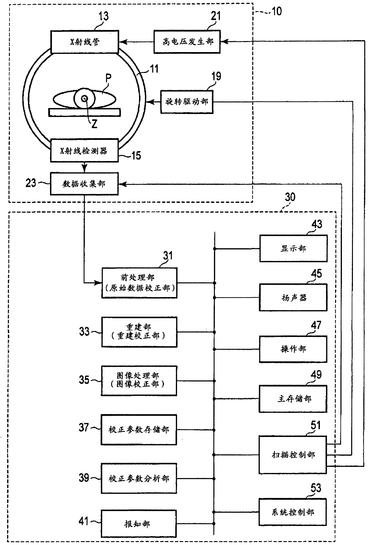
[0012] figure 1 It is a figure which shows the structure of the X-ray computed tomography apparatus concerning this embodiment. like figure 1 As shown, the X-ray computed tomography apparatus according to this embodiment has a gantry 10 and a console 30 . The gantry 10 is installed in a CT imaging room such as a hospital. The console 30 is installed in a CT imaging room, a control room adjacent to the CT imaging room, or the like. The console 30 controls the gantry 10 in accordance with instructions and the like from an operator.

[0013] The stand 10 is equipped with a rotating frame 11 in a casing (not shown) formed with an opening. The rotating frame 11 is accommodated in the casing so that the central axis Z of the casing coincides with the central axis (rotation axis) Z of the rotati...


PUM


Abstract

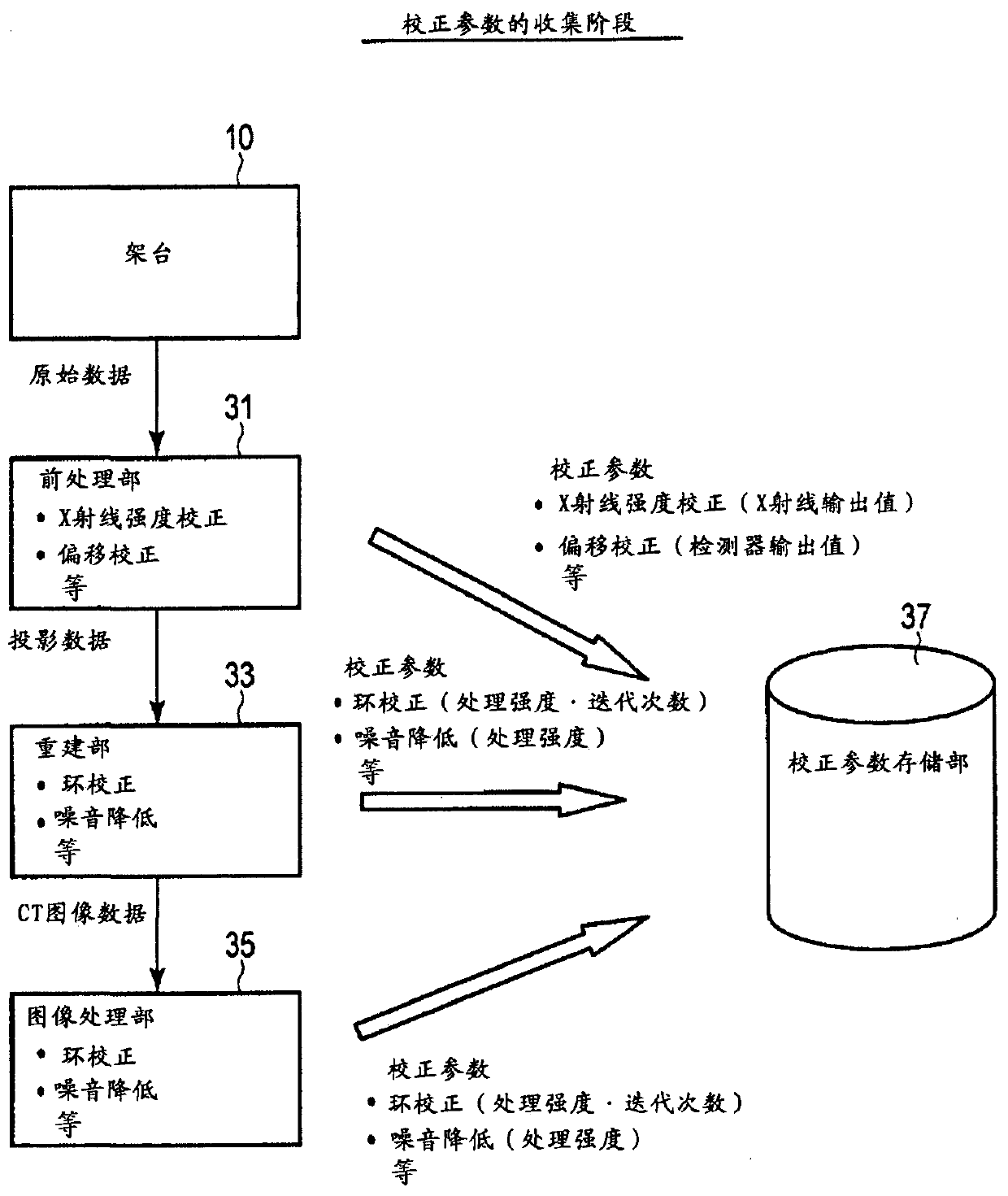
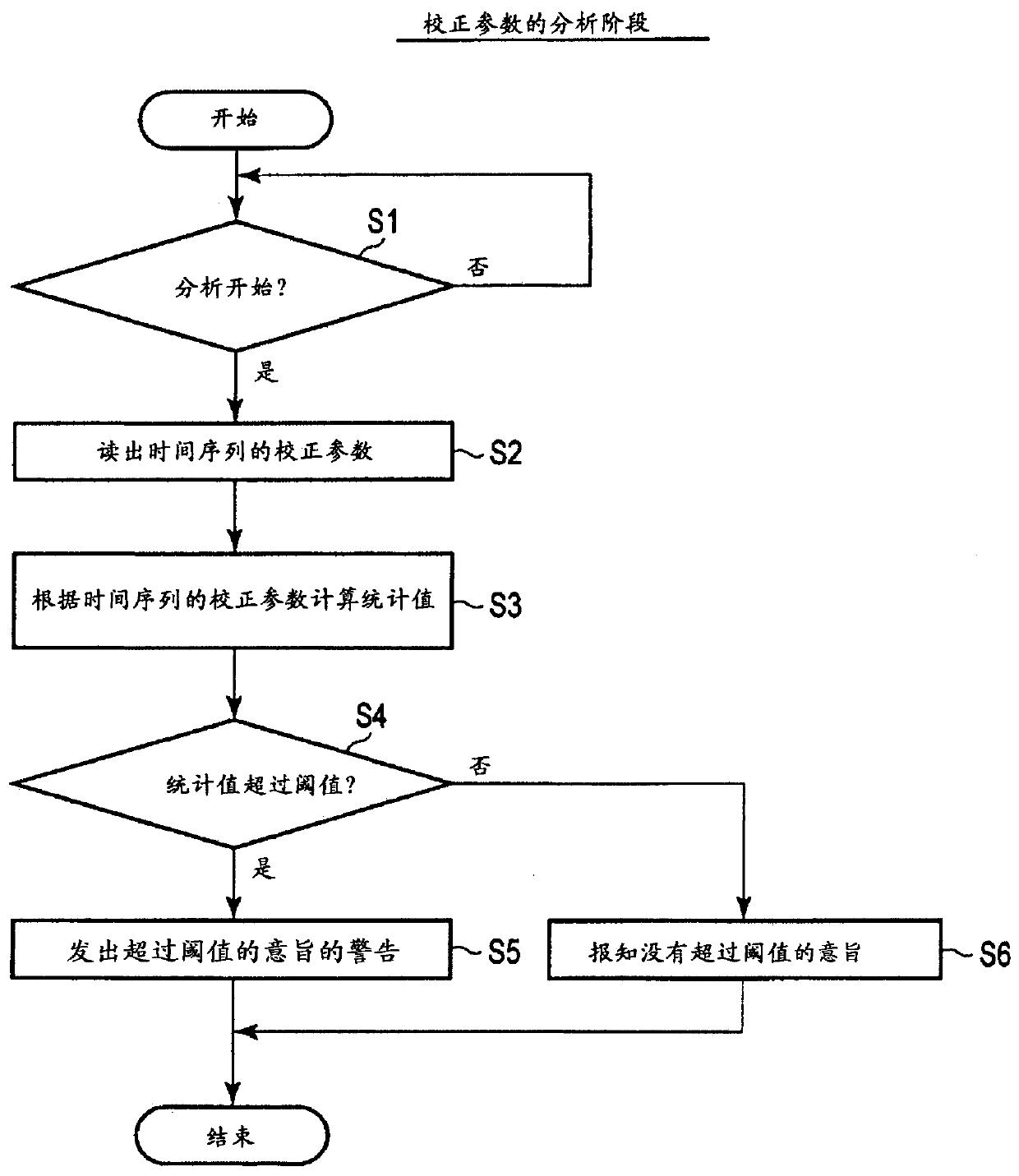
Grasp the working status of the device at any point in time. The reconstructed image generation unit (preprocessing unit 31 , reconstruction unit 33 , image processing unit 35 ) generates a reconstructed image based on output data based on electrical signals from the X-ray detector 15 . The correction parameter storage unit 37 stores correction parameters related to correction performed to reduce noise or artifacts in time series during the process of the reconstructed image generation unit. The correction parameter analysis unit 39 determines whether or not an abnormality has occurred based on temporal changes in the correction parameters stored in the correction parameter storage unit 37 . When it is determined by the correction parameter analysis unit 39 that an abnormality has occurred, the notification unit 41 notifies that the abnormality has occurred.

Description

technical field [0001] This embodiment relates to an X-ray computed tomography apparatus and an information processing apparatus. Background technique [0002] Currently, abnormal states of X-ray computed tomography apparatuses are often detected. For example, an abnormality of an X-ray computed tomography apparatus can be confirmed by observing an error log or reconstructing an image. At this time, the change in the state of the device cannot be noticed unless it is in an abnormal state. In addition, there is also a method of detecting an abnormal state by performing a CT scan on a phantom for image quality analysis at the time of periodic maintenance or the like. In this method, an abnormality is detected by checking the output of an X-ray detector by a service person or the like, reconstructing an image, or automatically analyzing by analysis software. In this case, only periodic confirmation is possible. Contents of the invention [0003] An object of the embodimen...

Claims


Application Information

Patent Timeline
Patent Type & Authority Patents(China)
IPC IPC(8): A61B6/03
CPCA61B6/5258A61B6/586A61B6/032A61B6/5205A61B6/5264A61B6/563G06T7/0012G06T2207/10004G06T2207/10081
Inventor 山鼻将央川锅信哉养田隆宏大西莉纱小贯广行
Owner TOSHIBA MEDICAL SYST CORP
Why Eureka
Eureka Blog
Learn More