Coronary artery automatic extraction method based on three-dimensional morphology

An automatic extraction and morphological technology, used in image analysis, image enhancement, instrumentation, etc., can solve problems such as high time complexity, low precision, and easy generation of outliers.

Active Publication Date: 2015-12-02
CHONGQING UNIV +1
View PDF3 Cites 31 Cited by
  • Summary
  • Abstract
  • Description
  • Claims
  • Application Information

AI Technical Summary

Problems solved by technology

[0003] Among these methods, the tracking-based method has too much manual intervention and high time complexity, while other methods have too high requirements on the strength of the image threshold, which is prone to outliers and has low precision.

Method used


Image

  • Coronary artery automatic extraction method based on three-dimensional morphology
  • Coronary artery automatic extraction method based on three-dimensional morphology
  • Coronary artery automatic extraction method based on three-dimensional morphology

Examples


Embodiment Construction

[0045] Embodiments of the present invention are described in detail below, examples of which are shown in the drawings, wherein the same or similar reference numerals designate the same or similar elements or elements having the same or similar functions throughout. The embodiments described below by referring to the figures are exemplary only for explaining the present invention and should not be construed as limiting the present invention.

[0046]In describing the present invention, it should be understood that the terms "longitudinal", "transverse", "upper", "lower", "front", "rear", "left", "right", "vertical", The orientation or positional relationship indicated by "horizontal", "top", "bottom", "inner", "outer", etc. are based on the orientation or positional relationship shown in the drawings, and are only for the convenience of describing the present invention and simplifying the description, rather than Nothing indicating or implying that a referenced device or eleme...


PUM


Abstract

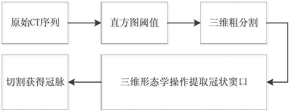
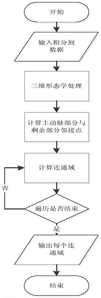
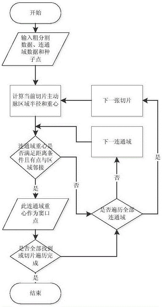
The present invention discloses a coronary artery automatic extraction method based on three-dimensional morphology. The method comprises the steps of (S1) obtaining image data, sorting images orderly according to obtainment times, and carrying out histogram transformation on the sorted images, (S2) carrying out three-dimensional coarse segmentation on a transformed image, carrying out three-dimensional morphology processing on the gray scale value of a split coronary artery image, and obtaining a communication domain formed by neighbor points through calculating the neighbor points of an aorta area and a corroded area, and (S3) traversing all neighbor points, after execution is completed, outputting the communication domain of each neighbor point, executing the process of cutting a coronary artery image, searching the opening point of the coronary artery from the inputted three-dimensional coarse segmentation data and the communication domain data of all neighbor points, and completing cutting of the coronary artery image.

Description

technical field [0001] The invention relates to the field of medical image extraction, in particular to an automatic extraction method for coronary vessels based on three-dimensional morphology. Background technique [0002] By extracting coronary vessels from CT images and analyzing the parameters and motion information of the sequences, abnormalities can be found, which is of great significance for revealing cardiovascular diseases. Accurately locate the spatial position of the lesion, and can be applied to virtual planning and surgical navigation to improve the quality and success rate of clinical operations. Cardiovascular segmentation is the basis for follow-up operations such as 3D reconstruction and quantitative analysis of normal and diseased tissues, and it is also the bottleneck of clinical medical applications. The accuracy of segmentation is crucial for doctors to judge the real situation of the disease and make a correct diagnosis plan. After years of developm...

Claims


Application Information

Patent Timeline
Patent Type & Authority Applications(China)
IPC IPC(8): G06T7/00
CPCG06T7/0012G06T2207/10081G06T2207/30101
Inventor 王翊房斌欧阳自鹏张绍祥谭立文李颖
Owner CHONGQING UNIV
Why Eureka
Try Eureka