A stress detection device around an intracranial hematoma in SD rats
A technology for intracranial hematoma and stress detection, applied in application, diagnostic recording/measurement, medical science, etc.
- Summary
- Abstract
- Description
- Claims
- Application Information
AI Technical Summary
Problems solved by technology
Method used
Image
Examples
Embodiment 1
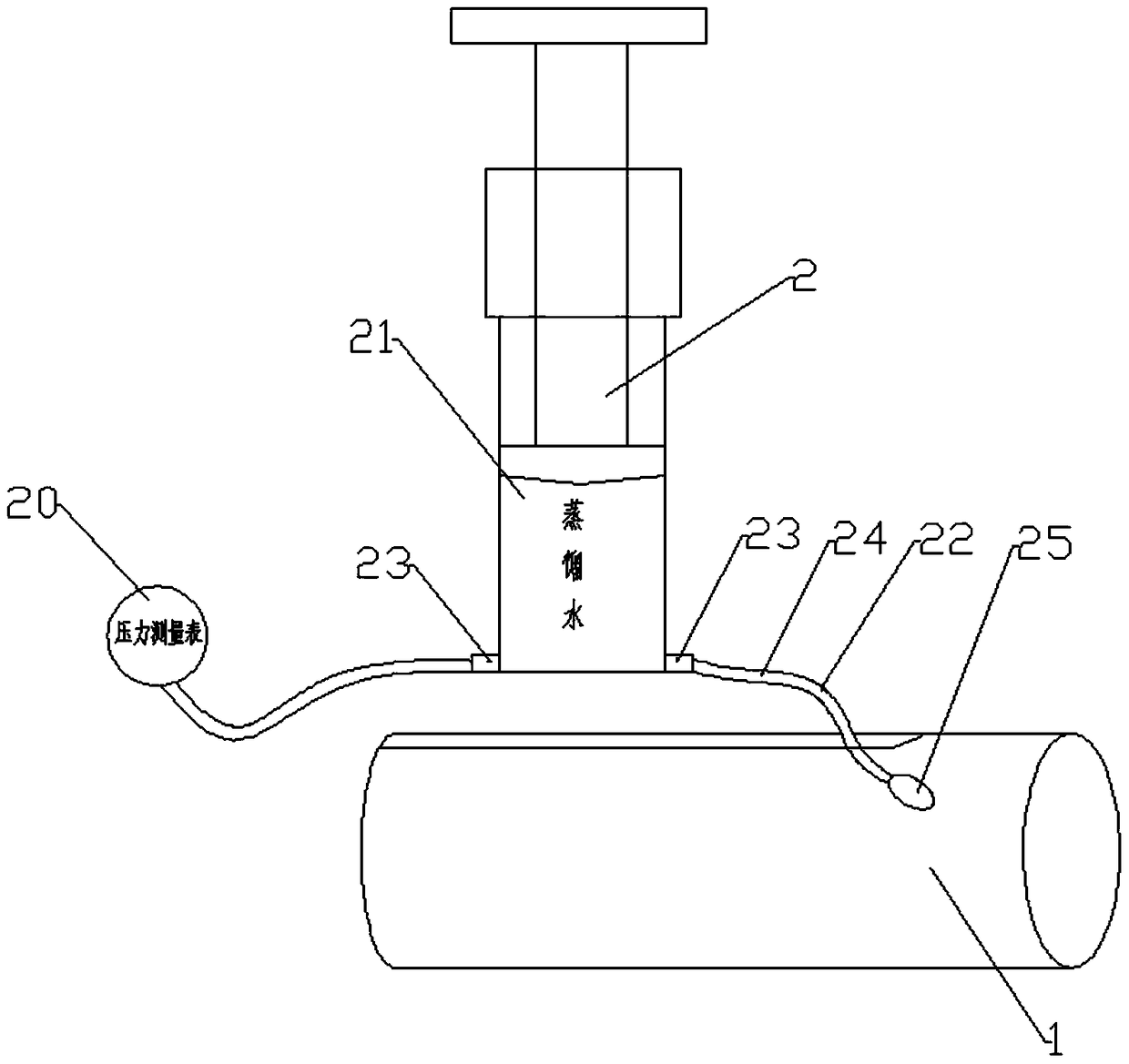
[0029] Embodiment 1: as figure 1 and Figure 5 As shown, an animal intracranial stress detection device includes a fixing device 1 for fixing the animal and a measuring device 2 for measuring the intracranial pressure of the animal. The measuring device 2 includes a pressure measuring gauge 20, a syringe 21 and a pressure device 22 connected to the intracranial of the animal. Two horizontally facing outlets 23 are arranged at the front end of the syringe 21, and one of the outlets 23 is connected to the The pressure measuring gauge 20, another outlet 23 is connected to the pressure applying device 22. Wherein, the pressure applying device 22 includes a catheter 24, one end of the catheter 24 is connected to the outlet 23 of the syringe 21, and the other end of the catheter 24 is provided with a balloon 25 placed in the animal's skull. The balloon 25 is fixedly connected to the skull of the animal. And a depth indicator line 28 is provided on the catheter at the upper end of...
Embodiment 2
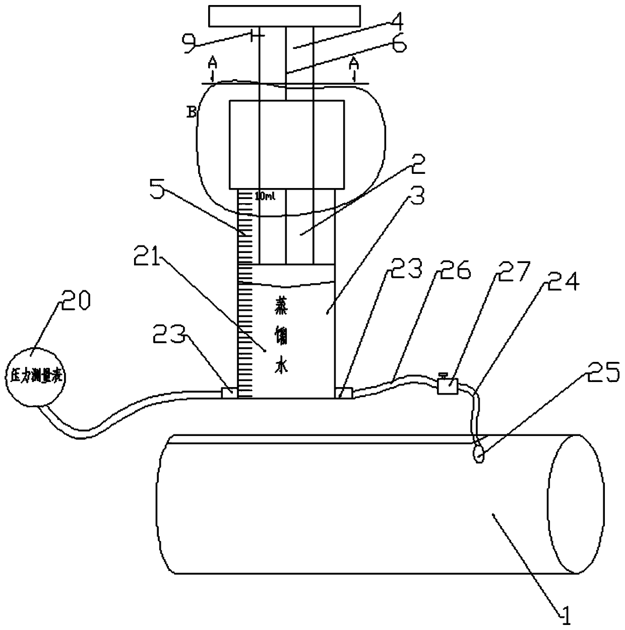
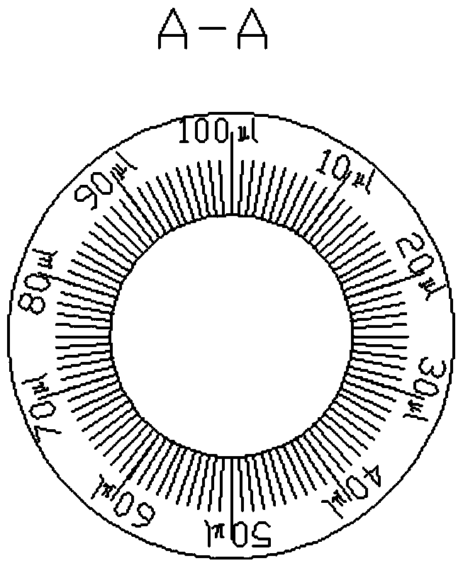
[0033] Embodiment 2: as Figure 2-5 As shown, an animal intracranial stress detection device includes a fixation device 1 for fixing the animal and a measuring device 2 for measuring the intracranial pressure of the animal. The measuring device 2 includes a pressure measuring gauge 20, a syringe 21 and a connecting animal The intracranial pressure applying device 22 is provided with two horizontally facing outlets 23 at the front end of the syringe 21 , and one of the outlets 23 is connected to the pressure measuring gauge 20 through a catheter (length: 40mm). Another outlet 23 is connected to the pressure applying device through a connecting pipe 26, and the pressure applying device includes a conduit 24, one end of the conduit 24 is connected with the connecting pipe 26, and the other end of the conduit 24 is provided with a An intracranial balloon 25, said balloon 25 is fixedly connected on the animal skull (around the hematoma), and a depth indicator line 28 is arranged on t...
Embodiment 3
[0039] Embodiment 3: as Figure 6 and Figure 7 As shown, this embodiment is an improvement on the fixing device 1 on the basis of Embodiment 1 and Embodiment 2. In this embodiment, the fixing device 1 includes a cylinder 10, a head baffle 11 and a tail The baffle plate 12 is provided with an axial chute 14 on the cylinder 10, the chute 14 has an opening at one end and a closed end at the other end, a fixer 15 is provided on the head baffle, and the The head baffle 11 is put in from the end of the cylinder 10, the fixer 15 is exposed through the chute 14, and slides into the interior of the cylinder 10 through the chute 14 until it is put into the chute 14. Close the end, then put the animal to be detected into the cylinder 10, and finally put the tail baffle 12 with the holder 15 into the cylinder 10 in the same way.
[0040] In addition, an opening 13 capable of exposing the tail of the animal is also provided on the tail baffle 12 , so that it is convenient to perform mer...
PUM
 Abstract
Description
Claims
Application Information
 - R&D
- Intellectual Property
- Life Sciences
- Materials
- Tech Scout
- Unparalleled Data Quality
- Higher Quality Content
- 60% Fewer Hallucinations
Browse by: Latest US Patents, China's latest patents, Technical Efficacy Thesaurus, Application Domain, Technology Topic, Popular Technical Reports.
© 2025 PatSnap. All rights reserved.Legal|Privacy policy|Modern Slavery Act Transparency Statement|Sitemap|About US| Contact US: help@patsnap.com


