Ultrasound three-dimensional imaging control method, imaging method and system

A three-dimensional imaging and control method technology, applied in ultrasonic/sonic/infrasonic diagnosis, sonic diagnosis, infrasonic diagnosis, etc., can solve the problems affecting the diagnosis results, image resolution, low image resolution, low image resolution, information loss, etc. Effects on the probability of misdiagnosis and missed diagnosis

Active Publication Date: 2017-12-12
SONOSCAPE MEDICAL CORP
View PDF7 Cites 0 Cited by
  • Summary
  • Abstract
  • Description
  • Claims
  • Application Information

AI Technical Summary

Problems solved by technology

However, due to the insufficient amount of original volume data, this kind of 3D image has the defects of missing information, low image resolution and low slice image resolution, which affects the diagnosis results and easily leads to misdiagnosis and missed diagnosis by clinicians.

Method used


Image

  • Ultrasound three-dimensional imaging control method, imaging method and system
  • Ultrasound three-dimensional imaging control method, imaging method and system
  • Ultrasound three-dimensional imaging control method, imaging method and system

Examples


Embodiment 1

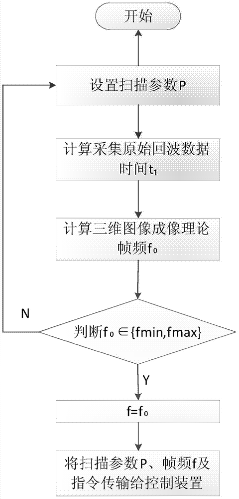
[0027] This embodiment provides a method for controlling ultrasonic three-dimensional imaging, the method comprising:

[0028] S1. The ultrasound host sets and adjusts the scanning parameter P;

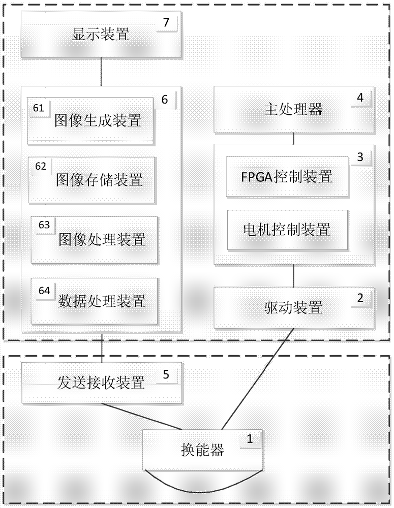
[0029] like figure 1 As shown, in the three-dimensional ultrasonic imaging system, the ultrasonic probe includes at least a transducer 1, a motor, and a transmission mechanism (not shown), and the system host includes at least a driving device 2, a control device 3 and a main processor that are sequentially connected in communication with the motor 4. The main processor 4 can set and adjust the scanning angle θ according to requirements 1 , scanning depth D, single-frame grayscale image line number N, single-frame grayscale image opening angle θ 2 Wait for the scanning parameter P, and can send the scanning parameter P and other instructions to the control device 3 including FPGA control and scanning control, wherein the three-dimensional image imaging frame frequency f value and t...

Embodiment 2

[0039] The ultrasonic three-dimensional imaging control method provided in this embodiment includes:

[0040] S1. Set and adjust the scanning parameter P of the ultrasound host computer; figure 2 shown, including,

[0041] S11. Set the scanning parameter P, at least including the scanning angle θ 1 , scanning depth D, single-frame grayscale image scanning line number N, single-frame grayscale image opening angle θ 2 , adjustment coefficient K;

[0042] S12. Calculate the original echo data acquisition time t 1 ;

[0043] For example, in order to ensure that the scanning line data density of the two-dimensional grayscale image in the scanning direction of the ultrasonic probe transducer is the same as that of the X-section of the three-dimensional image, set the scanning parameter P and the acquisition time t 1 functional relationship.

[0044] In this embodiment, the acquisition time t is calculated according to the following formula 1 : C is the average propagation ...

Embodiment 3

[0063] This embodiment provides an ultrasonic three-dimensional imaging method, including:

[0064] S1. The ultrasound host sets and adjusts the scanning parameter P;

[0065] The main processor 4 can set and adjust the scanning angle θ according to requirements 1 , scanning depth D, single-frame grayscale image line number N, single-frame grayscale image opening angle θ 2 Wait for the scanning parameter P, and can send the scanning parameter P and other instructions to the control device 3 including FPGA control and scanning control, wherein the three-dimensional image imaging frame frequency f value and the scanning parameter P are functional relationships.

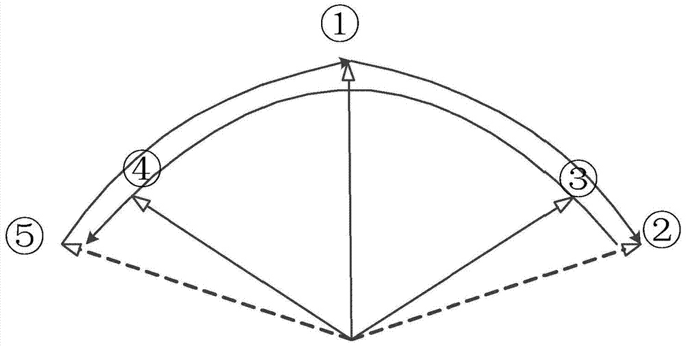
[0066] S2. Control the scanning of the three-dimensional ultrasonic probe transducer 1 according to the scanning parameter P;

[0067] The control device 3 controls the signal output by the driving device 2, and then enables the motor and the transmission mechanism to drive the transducer 1 to perform logical trajecto...


PUM


Abstract

The invention provides a method for controlling ultrasonic three-dimensional imaging, comprising: setting and adjusting the scanning parameter P by the ultrasonic host computer; wherein, the frame frequency f value of the three-dimensional image imaging and the scanning parameter P are functional relationships; controlling the scanning parameter P according to the Scanning with a three-dimensional ultrasonic probe transducer; the ultrasonic probe transducer collects original echo data of a two-dimensional grayscale image; processes the original echo data of the two-dimensional grayscale image to construct a three-dimensional image and / or at least one section image . The method provided by the invention designs the three-dimensional image imaging frame frequency f value and the scanning parameter P as a function relationship, which can realize three-dimensional images with low frame frequency and high image resolution, and is especially suitable for static tissues, such as breast, thyroid, and pelvis. Fundus, uterus, and accessories do not require high real-time 3D images and focus on image resolution imaging of tissue parts, reducing the probability of misdiagnosis and missed diagnosis by clinicians due to low image resolution affecting diagnostic results.

Description

technical field [0001] The invention relates to the field of ultrasonic imaging, in particular to an ultrasonic three-dimensional imaging control method, imaging method and system. Background technique [0002] The three-dimensional imaging technology of the ultrasound imaging system has always been a research and development hotspot in this field because it can present the scanned object three-dimensionally and facilitate doctors to diagnose and treat the scanned object. Ultrasound three-dimensional imaging is a three-dimensional data structure transformed by computer processing on the basis of a series of continuous two-dimensional plane data collected. Due to the limited amount of original 2D echo data collection and transmission speed, in order to ensure the real-time performance of 3D image acquisition, the number of lines of each frame of raw data and the number of frames of scanned volume data are usually reduced. When the amount of original volume data is insufficie...

Claims


Application Information

Patent Timeline
Patent Type & Authority Patents(China)
IPC IPC(8): A61B8/00G06F19/00
Inventor 李萍陈维楚黄柳倩艾金钦唐艳红
Owner SONOSCAPE MEDICAL CORP
Why Eureka
Try Eureka