Breast volume ultrasound imaging device and method

An ultrasonic imaging method and ultrasonic imaging technology, applied in the directions of ultrasonic/sonic/infrasonic image/data processing, ultrasonic/sonic/infrasonic Permian technology, organ movement/change detection, etc. Unfavorable follow-up observation, inaccurate estimation of tumor shape and size, etc., to achieve the effect of enhancing repeatability and facilitating inspection and diagnosis

Active Publication Date: 2018-03-23
NANJING STARTON MEDICAL TECH CO LTD
View PDF7 Cites 0 Cited by
  • Summary
  • Abstract
  • Description
  • Claims
  • Application Information

AI Technical Summary

Problems solved by technology

Clinically, doctors need to spell out the three-dimensional structure of breast tissue in their minds based on real-time two-dimensional images, which requires doctors to have very rich clinical experience and long-term training, which is easy to produce inaccurate information about the location, shape and size of tumors In the follow-up ultrasound examination, according to the previous two-dimensional ultrasound image, it is difficult for the doctor to locate the image obtained by the previous examination, and it is also difficult to obtain the ultrasound image of the same position repeatedly, which is very important for the long-term follow-up observation of the lesion. Adverse

Method used


Image

  • Breast volume ultrasound imaging device and method
  • Breast volume ultrasound imaging device and method
  • Breast volume ultrasound imaging device and method

Examples


Embodiment Construction

[0034] In order to better understand the technical content of the present invention, specific embodiments are given together with the attached drawings for description as follows.

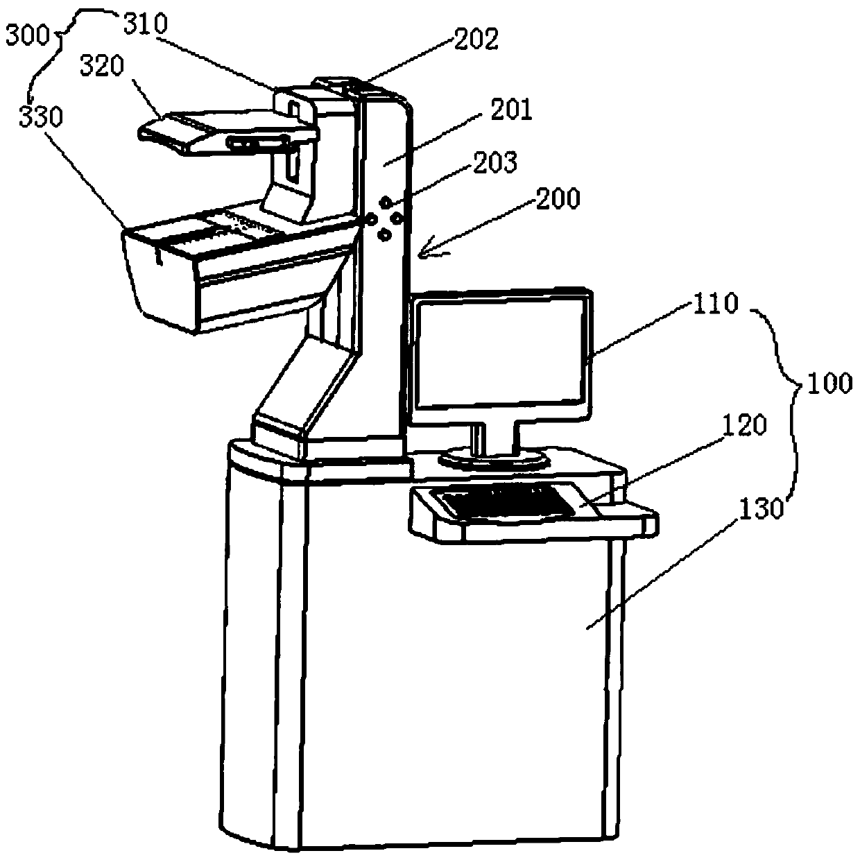
[0035] Such as figure 1 As shown, according to a preferred embodiment of the present invention, a breast volume ultrasound imaging device includes a main body 100, a main frame 200 and a probe mechanism 300, the main frame 200 is located on the upper part of the main body 100 for providing The probe mechanism 300 is supported, and the probe mechanism 300 can be operated and controlled from the outside to move or turn over.

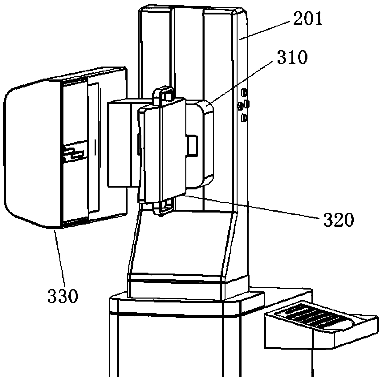
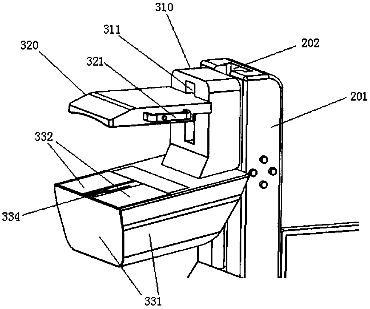
[0036] Such as figure 2 As shown, the aforementioned probe holder 310, detection and scanning assembly 330, and the detection pressing mechanism 320 installed on the probe holder 310 adopt a reversible design, for example image 3 As shown, the probe holder 310 , the detection and scanning assembly 330 and the detection and compression mechanism 320 are turned over as a whol...


PUM


Abstract

The invention provides a mammary gland volume ultrasonic imaging device and method, which realize three-dimensional volume ultrasonic imaging of mammary glands, so as to facilitate the examination and diagnosis of mammary gland diseases. The device consists of a main body, a main frame and a probe mechanism, which can move up and down and / or rotate for vertical or horizontal detection. The probe mechanism consists of a detection bracket, a pressing and fixing device, and a detection and scanning mechanism. The breast tissue to be tested is clamped in the middle, the upper clamping device can move up and down, and the lower scanning device has an ultrasonic probe and a moving mechanism. During one imaging process, the ultrasonic probe scans the tested breast and obtains The position data of the ultrasonic probe and ultrasonic sampling images at each moment, and then the ultrasonic slice image group with position information obtained through image processing, and three-dimensional image reconstruction is performed based on this, to obtain three-dimensional ultrasonic volume imaging of breast tissue.

Description

technical field [0001] The invention relates to the technical field of ultrasonic diagnosis, especially breast ultrasonic scanning, medical image processing and reconstruction, in particular to a breast volume ultrasonic imaging device and method. Background technique [0002] Mammography is a commonly used breast imaging detection method in the prior art. Imaging is simple and reproducible, and is currently the gold standard for breast cancer detection. However, mammography has X-ray radiation, requires a special protective environment, and is not effective for dense breast examination. Studies have shown that for dense breast tissue, mammography reports a sensitivity of 44%. dense breasts (i.e. BI-RADS TM Breast density of 3 or 4) is common in young women, while compared with Western women, Eastern women have relatively dense breasts, and the age of onset of breast cancer is lower. So other testing equipment is needed. [0003] In addition, magnetic resonance imaging ...

Claims


Application Information

Patent Timeline
Patent Type & Authority Patents(China)
IPC IPC(8): A61B8/08
Inventor 王维平
Owner NANJING STARTON MEDICAL TECH CO LTD
Why Eureka
Try Eureka