Thoracic contour data acquisition device for electrical impedance tomography of human thoracic cavity

A technology of tomography and data acquisition, applied in applications, medical science, sensors, etc., can solve problems such as outdated time

Inactive Publication Date: 2016-05-04
TIANJIN UNIV
View PDF4 Cites 3 Cited by
  • Summary
  • Abstract
  • Description
  • Claims
  • Application Information

AI Technical Summary

Problems solved by technology

Therefore, it is out of date to directly use the existing EIT da

Method used


Image

  • Thoracic contour data acquisition device for electrical impedance tomography of human thoracic cavity
  • Thoracic contour data acquisition device for electrical impedance tomography of human thoracic cavity
  • Thoracic contour data acquisition device for electrical impedance tomography of human thoracic cavity

Examples


Embodiment Construction

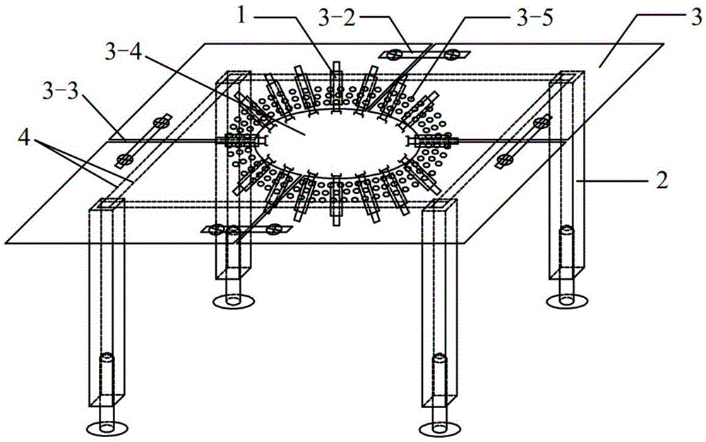
[0019] The structure and the functions of each part of a chest cavity contour data acquisition device for human chest cavity electrical impedance tomography of the present invention are described in conjunction with the accompanying drawings:

[0020] Such as figure 1 As shown, the data acquisition device includes a displacement measurement sensor 1 , a bracket 2 , a support panel 3 and a load-bearing beam 4 and other parts. The displacement measurement sensor 1 is arranged on the support panel 3, through a number of via holes 3-5 on the support panel 3, the angle and position between it and the chest cavity of the human body can be adjusted arbitrarily, after the adjustment is completed, the via holes are passed through with screws 3-5 Fix the displacement measuring sensor 1 on the support panel 3 . The displacement measurement sensor 1 is used to measure the position information of the human chest cavity in real time. The end close to the human chest cavity can be buckled w...


PUM


Abstract

The invention provides a thoracic contour data acquisition device for electrical impedance tomography of a human thoracic cavity. The data acquisition device comprises the following parts: a plurality of displacement measurement sensors (1), a bracket (2), a supporting panel (3) and the like, wherein the supporting panel (3) is fixedly arranged on the bracket (2); a hole (3-4), which can accommodate the human thoracic cavity, is formed in the middle of the supporting panel (3); a plurality of through holes (3-5) are formed around the hole (3-4) of the supporting panel (3); each of the displacement measurement sensor (1) is fixedly connected to the supporting panel (3) by virtue of at least two through holes (3-5); in accordance with the various positions of the corresponding through holes (3-5), the included angles and the positions of the displacement measurement sensors (1) relative to the human thoracic cavity are regulated; and the displacement measurement sensor (1) is used for measuring the position information of the human thoracic cavity in real time. By virtue of the data acquisition device disclosed by the invention, the spatial position information required by an updated field domain model can be acquired in real time.

Description

[0001] Technical Field The present invention belongs to the technical field of bioelectrical impedance tomography, and in particular relates to a chest contour data acquisition device for human chest electric impedance tomography. Background technique [0002] Electrical impedance tomography (Electrical Impedance Tomography, EIT) technology is a new type of non-invasive biomedical detection and imaging technology that uses the distribution or change of electrical impedance in the organism as the imaging target. The impedance distribution inside the object to be measured reflects the biological characteristics of each biological tissue structurally and functionally. EIT technology is a new type of process detection technology, which has shown great application prospects in medical image-based diagnostic technology. Using EIT technology, it is possible to display the impedance distribution image of the tissue in the living body, the image of the impedance change of the biologica...

Claims


Application Information

Patent Timeline
IPC IPC(8): A61B5/053A61B5/113
CPCA61B5/0536A61B5/113
Inventor 岳士弘郝振华
Owner TIANJIN UNIV
Why Eureka
Try Eureka