Method and system for respiratory monitoring during ct-guided interventional procedures

A respiratory monitoring system and respiratory monitoring technology, which are used in surgical navigation systems, instruments for radiological diagnosis, and surgical system user interfaces, etc. question

Inactive Publication Date: 2016-08-03
KONINKLJIJKE PHILIPS NV
View PDF7 Cites 2 Cited by
  • Summary
  • Abstract
  • Description
  • Claims
  • Application Information

AI Technical Summary

Problems solved by technology

However, this method has significant drawbacks: the diaphragm position between breath stops is not reproducible; and after a breath hold, subsequent deep breaths may cause instrumental injury to the patient, e.g., puncture of the pleura
Also, some patients (especially those with lung problems) may have difficulty holding their breath

Method used


Image

  • Method and system for respiratory monitoring during ct-guided interventional procedures
  • Method and system for respiratory monitoring during ct-guided interventional procedures
  • Method and system for respiratory monitoring during ct-guided interventional procedures

Examples


Embodiment Construction

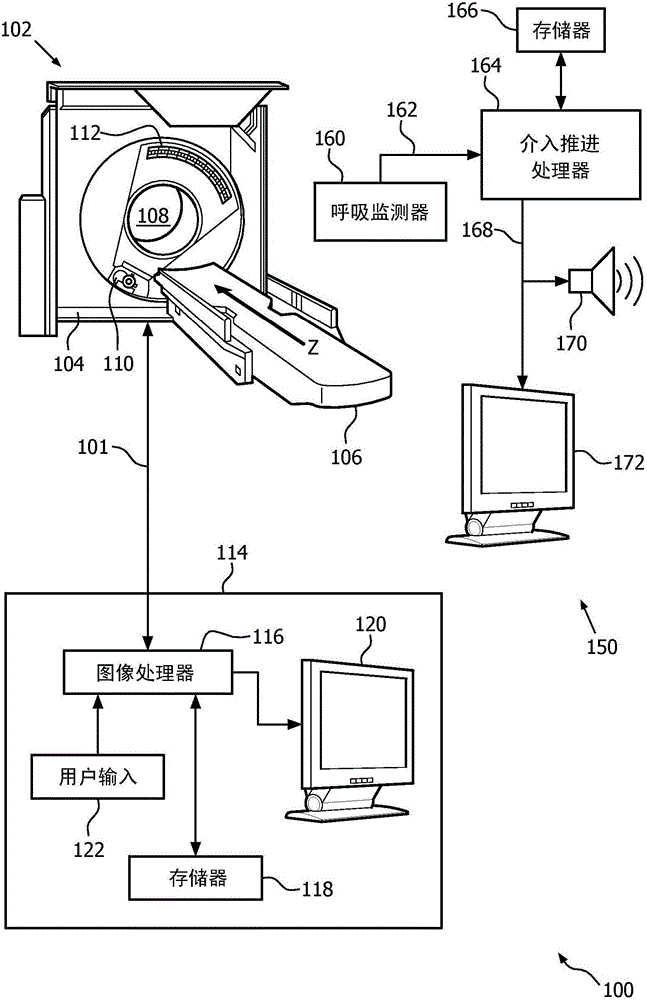
[0025] In one embodiment, figure 1 An exemplary CT imaging system 100 and an exemplary interventional tool advancement indicator system 150 are shown. The CT imaging acquisition system 102 includes a gantry 104 and a table or other support 106 movable along the z-axis. The patient or other object to be imaged ( figure 1 not shown) lies on the table 106 and is moved to be disposed within the aperture or bore 108 of the gantry 104. Once the patient is in position, the x-ray source 110 and x-ray detector 112 are rotated together about the bore 108 to record CT imaging data. Other imaging system modalities may also be used in conjunction with the claimed invention including, for example, cone beam CT, other X-ray based imaging, ultrasound imaging, magnetic resonance imaging (MRI), positron emission tomography (PET )Wait.

[0026] Afterwards, the CT imaging acquisition system 102 transmits the CT imaging data to the CT imaging processing and display system 114 through the commu...


PUM


Abstract

A method and system for respiratory monitoring and indicating intervention tool advancement timing during an intervention procedure. Features include monitoring the respiration of a subject using a respiration monitor, wherein the respiration monitor produces a respiratory signal indicative of a plurality of respiratory states of the subject, scanning the subject using an imaging device and generating a first imaging data set, wherein the imaging data set is associated with a respiratory state of the subject, and indicating when to advance an intervention tool based on the respiratory signal using an advancement indicator, such that advancement of the intervention tool occurs during the respiratory state of the subject, and where the subject need not hold his breath.

Description

technical field [0001] The present application generally relates to a method and system for breath-based guidance during interventional procedures. Interventional procedures can include interactive image-guided surgery and interactive surgical procedures, eg, biopsies. It finds particular application in minimally invasive surgical procedures performed in conjunction with x-ray computed tomography (CT) imaging systems. These procedures involve the use of biopsy surgical tools or brachytherapy needles, etc., to take tissue samples or to plan or place objects or instruments inside a subject, such as a patient. However, it should also be appreciated that the present invention is also applicable to a wide range of other imaging devices and techniques, for example, ultrasound and magnetic resonance imaging devices, PET, SPECT, etc., and to a wide range of minimally invasive surgical procedures, including Many forms of surgery that focus on placing objects or instruments at precise...

Claims


Application Information

Patent Timeline
Patent Type & Authority Applications(China)
IPC IPC(8): A61B5/113A61B6/03A61B6/00A61B5/00A61B5/055A61B6/12
CPCA61B5/7285A61B6/032A61B6/037A61B6/12A61B6/486A61B6/5288A61B6/541A61B5/055A61B5/1135A61B2505/05A61B2017/00699A61B34/10A61B34/20A61B2034/2046A61B34/25A61B2034/252A61B5/091A61B5/6804A61B6/4435
Inventor S·戈特曼
Owner KONINKLJIJKE PHILIPS NV
Why Eureka
Try Eureka