Check patentability & draft patents in minutes with Patsnap Eureka AI!

Multi-atlas dividing method for low-resolution medical image

A low-resolution image and high-resolution image technology, applied in the field of multi-spectrum segmentation of low-resolution medical images, can solve the problems of unguaranteed target image quality, low-resolution target images, etc., and improve the segmentation accuracy is not high , Improve the effect of segmentation accuracy

Inactive Publication Date: 2016-08-31
SHAOXING UNIVERSITY
View PDF4 Cites 14 Cited by
  • Summary
  • Abstract
  • Description
  • Claims
  • Application Information

AI Technical Summary

Problems solved by technology

[0005] In the practical application of the multi-atlas segmentation method, since the atlas is pre-constructed, high-resolution image construction is usually used to obtain a high-quality atlas; however, affected by factors such as image acquisition time and acquisition equipment, the quality of the target image cannot be guaranteed. will get a low-resolution target image

Method used


Image

  • Multi-atlas dividing method for low-resolution medical image
  • Multi-atlas dividing method for low-resolution medical image
  • Multi-atlas dividing method for low-resolution medical image

Examples


Embodiment Construction

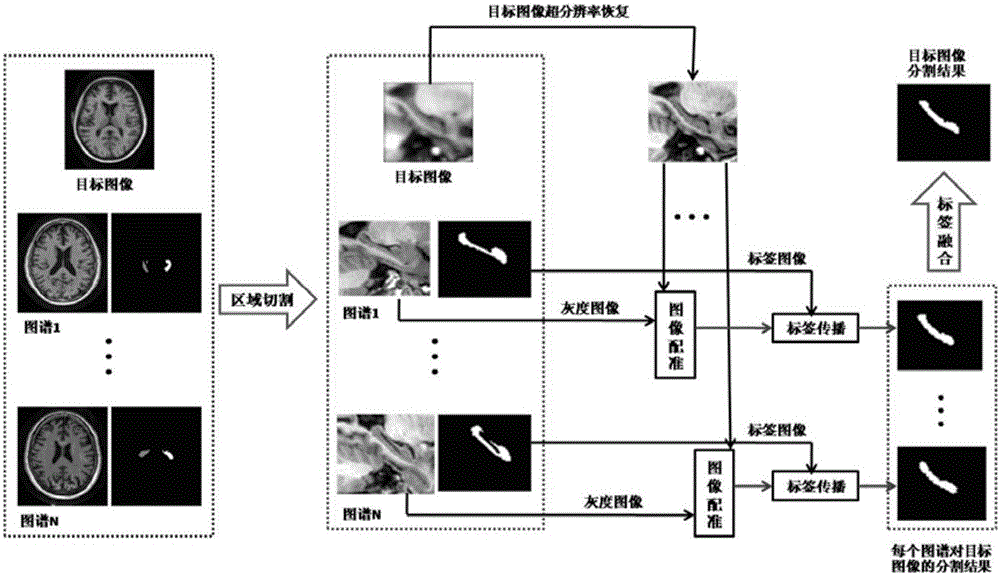
[0031] A multi-atlas segmentation method for low-resolution medical images, comprising the following steps:

[0032] Step 1. Given a low-resolution target image I d , N high-resolution atlas images A i =(I i , L i ), i=1,2,...,N, where I iIndicates the i-th grayscale image, L i Indicates the label image corresponding to the i-th grayscale image, and assumes the target image I d and atlas image A i =(I i , L i ), i=1,2,...,N, have been linearly registered to the same template space;

[0033] Step 2. Segment object area cutting:

[0034] Such as figure 1 As shown, in order to reduce the computational workload, a region containing the segmentation object is cut off first. Since the target image I d and atlas image A i =(I i , L i ), i=1, 2,..., N, have been linearly registered to a template space, so the positions of the segmented objects in each image are roughly similar, scan all atlas images A i =(I i , L i ), i=1, 2,..., N, label images in (that is, scan all...


PUM


Abstract

The invention discloses a multi-atlas dividing method for a low-resolution medical image, wherein the multi-atlas dividing method belongs to the field of image processing technology. The method comprises the steps of setting a low-resolution objective image and N high-resolution atlas images, and assuming a fact that the objective image and the atlas images are linearly registered in a same template space; and then successively performing a dividing object area cutting step; an objective image super-resolution recovering step; an image registering and label propagation step; and a label fusion step. According to the multi-atlas dividing method for the low-resolution medical image, an image super-resolution restoring method is merged into a multi-atlas dividing frame; and through improving registering precision between the high-resolution atlas images and the low-resolution to-be-divided image, dividing precision of the multi-atlas dividing method.

Description

technical field [0001] The invention relates to a multi-atlas segmentation method of a low-resolution medical image, belonging to the technical field of image processing. Background technique [0002] With the development and popularization of medical imaging equipment, medical image analysis plays an extremely important role in disease research, surgical planning, and clinical diagnosis. Medical image segmentation is an important part of medical image analysis. Its essence is to label each pixel or voxel of the image to be segmented, and set different label values ​​for pixels or voxels with different attributes, so as to divide the image to be segmented into infinite Overlapping regions of interest and background regions for further analysis and processing of regions of interest. Usually, the segmentation of medical images is completed by manually marking the region of interest. The advantage of this method is that the accuracy of the segmentation results is high. Howeve...

Claims


Application Information

Patent Timeline
IPC IPC(8): G06T7/00G06T5/00
CPCG06T2207/30004G06T5/00
Inventor 祝汉灿范勇
Owner SHAOXING UNIVERSITY
Features
  • R&D
  • Intellectual Property
  • Life Sciences
  • Materials
  • Tech Scout
  • Unparalleled Data Quality
  • Higher Quality Content
  • 60% Fewer Hallucinations
Learn More