
Pathology identification method for routine scan CT image of liver based on random forests

A random forest algorithm and CT image technology, applied in the field of lesion recognition in plain CT images of the liver, can solve problems such as misdiagnosis, slow postoperative recovery, missed treatment time, etc., achieve great medical value and reduce the effect of error rate

Inactive Publication Date: 2016-09-07
ZHEJIANG UNIV
View PDF4 Cites 43 Cited by
  • Summary
  • Abstract
  • Description
  • Claims
  • Application Information

AI Technical Summary

Problems solved by technology

At present, liver cancer has become one of the diseases with the highest fatality rate in the world. Due to the lack of treatment methods and the lack of obvious pathological indicators of early liver cancer, it may cause misdiagnosis and miss the best time for treatment.
The diagnosis of liver cancer mainly relies on liver biopsy technology, but this technology will cause certain damage to the patient's liver, coupled with the high difficulty of implementation and slow postoperative recovery. Therefore, the current diagnosis of liver diseases mainly relies on medical imaging, such as liver CT

Method used


Image

  • Pathology identification method for routine scan CT image of liver based on random forests
  • Pathology identification method for routine scan CT image of liver based on random forests
  • Pathology identification method for routine scan CT image of liver based on random forests

Examples


Embodiment Construction

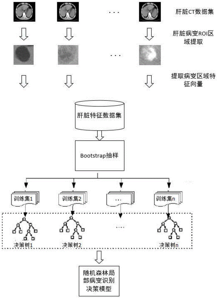
[0031] In order to realize the identification of local lesions in liver plain scan CT images of the present invention, the following two stages are adopted.

[0032] The first stage: establishment of lesion identification model based on random forest algorithm

[0033] 1. Establishment of image feature data set of local lesion area in plain CT image of liver:

[0034] The present invention uses 3,000 plain liver CT images marked from a hospital in Hangzhou, all of which are 512*512 in size, including several types such as normal, liver cancer, hepatic hemangioma, and liver cyst, etc., and extracts a rectangular frame that can cover the marked lesion area As the region of interest, 3000 lesion block images are obtained in this way.

[0035] For each lesion area block, calculate the gray histogram, gray level co-occurrence matrix and gray level gradient co-occurrence matrix of the lesion block image.

[0036] For the lesion block image, the calculation method of the gray histo...


PUM


Abstract

The invention discloses a pathology identification method for a routine scan CT image of the liver based on the random forests. The method comprises that image gray-level texture characteristic is extracted from a pathologic area of the routine scan CT image of the liver and serves as image characteristic vector expression, the random forests is used to select characteristics from the image characteristic vector of the pathologic area of the routine scan CT image of the liver to form a most effective characteristic combination, a most effective characteristic data set is trained and learned, the identification capability of a decision tree of random forests is balanced and optimized, and a final pathology identification model is obtained.

Description

technical field [0001] The invention relates to a method for identifying pathological changes in plain scan CT images of the liver based on a random forest algorithm, in particular to the introduction of the most effective feature selection method and the improvement of the random forest algorithm. Background technique [0002] With the development and maturity of medical imaging technology, medical imaging plays an important role in the diagnosis of liver diseases. At present, liver cancer has become one of the diseases with the highest fatality rate in the world. Due to the lack of treatment methods and the lack of obvious pathological indicators of early liver cancer, it may cause misdiagnosis and miss the best time for treatment. The diagnosis of liver cancer mainly relies on liver biopsy technology, but this technology will cause certain damage to the patient's liver, coupled with high difficulty in implementation and slow postoperative recovery. Therefore, the current ...

Claims


Application Information

Patent Timeline
Patent Type & Authority Applications(China)
IPC IPC(8): G06T7/00
CPCG06T7/0012G06T2207/10081G06T2207/20081G06T2207/30056
Inventor 金心宇武海涛金奇樑刘帆
Owner ZHEJIANG UNIV
Learn More