
Non-radioactive label immunoprecipitation method for detecting specific self-antibody MDA5 of inflammatory myopathy
An autoantibody and immunoprecipitation technology, applied in the direction of virus/bacteriophage, recombinant DNA technology, biochemical equipment and methods, etc., can solve the problems of high false positive rate, high cost of ELISA kit, etc., and achieve easy transfection and high efficiency. Expressive, increased sensitivity effects
- Summary
- Abstract
- Description
- Claims
- Application Information
AI Technical Summary
Problems solved by technology
Method used
Image

Examples
Embodiment 1
[0027] Example 1 Non-radiolabeled immunoprecipitation method for detecting inflammatory myopathy-specific autoantibody MDA5
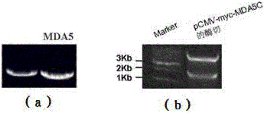
[0028] (1) Design primers MDA5-F (5'-TTCGGTCGACCAACAAAGAAGCAGTGTA-3') and MDA5-R (5'-GCGGTACCCTAATCCTCATCACTAAATAAACAG-3') to amplify the C-terminus of human MDA5 from the pENTER-MDA5 plasmid (Genbank: NM022168, 1743bp- 3479bp)( figure 1 a).
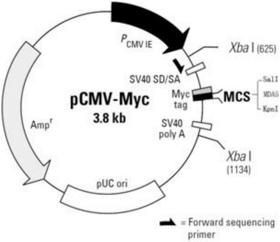
[0029] (2) Recover and purify the PCR fragment, perform double digestion with SalI and KpnI restriction endonucleases, connect overnight with the pCMV-myc vector that has been double-digested with SalI and KpnI, and transform JM109 bacteria competently, in the presence of 50ug / ml ampicillin Positive clones were screened on a penicillin plate, and the plasmid DNA of the positive clone was extracted in a small amount, and the plasmid DNA was digested with SalI and KpnI, and the results showed that the plasmid contained a 1.7kb insert ( figure 1 b).
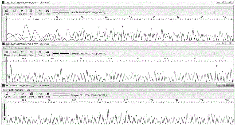
[0030] (3) The plasmid DNA is sequenced to confirm the accuracy...
Embodiment 2
[0033] Example 2 detects the positive rate of the MDA5 antibody in the serum of 50 patients with inflammatory myopathy
[0034] (1) Inoculate 30% HEK293 cells on 75cm 2 37 culture dishes for less than 24 hours;
[0035] (2) Transfect HEK293 cells with 30ug pCMV-myc-MDA5C plasmid for 48 hours;
[0036] (3) HEK293 cells were lysed with 1 ml of RIPA lysate containing protease inhibitors, and the protein was quantified;
[0037](4) The c-myc antibody was used to confirm the overexpressed MDA5 C-terminus in HEK293 cells.
[0038] Take 20ul of the test serum, positive serum, negative serum and cell lysate overexpressing the MDA5 C-terminus of patients with inflammatory myopathy for immunoprecipitation, perform SDS-PAGE gel electrophoresis, and then detect with c-myc antibody to determine the MDA5 antibody The positive rate in patients with inflammatory myopathy is about 10% ( Figure 6 For some patients with inflammatory myopathy detected).
PUM
 Abstract
Description
Claims
Application Information